Spirurids - Donkey
Stomach spirurid worms


Habronema muscae, H. majus and Draschia megastoma are frequently reported spirurids in donkeys (Pandey et al, 1994). Although the adults are considered to be non-pathogenic, the formation of large granulomas/tumours in the stomach by D. megastoma may interfere mechanically with its function. These lesions are frequent findings in the fundus region of the stomach of donkeys, often protruding into the lumen.
The life cycle involves an intermediate host, such as the stable fly, Stomoxys calicitrans, or the housefly, Musca domestica. The sprurid and the fly develop synchronously after the fly maggot has ingested larvae in the faeces. H. muscae and D. megastoma develop in the housefly, while H. majus develops in the stable fly. By the time the young fly is ready to seek a host the larvae become infective. The larvae are released onto the skin as the fly feeds and are ingested by the animal during grooming. Infection can also occur if the fly is swallowed. Only larvae gaining direct access to the stomach complete their development.
Diagnosis of stomach-worm infection is difficult because the eggs are not observed in routine faecal examination.
Cutaneous habronemiasis
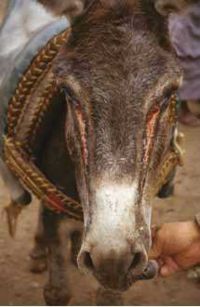
Cutaneous habronemiasis (summer sore) is a seasonal, granulomatous skin disease caused by aberrant Habronema larvae. It is caused by house and stable flies depositing infective larvae on moist mucous membranes or pre-existing skin lesions. Deposition of larvae in existing wounds is a major problem in the tropics among working donkeys. The problem is common during warm weather coinciding with the period of high fly activity. Body parts that commonly have wounds, moisture or discharges, i.e. lower limbs, back, girth and belly, mucocutaneous junctions such as sheath, prepuce, penis, lips, muzzle, eyelids and medial canthus of the eye, are common sites of lesions in donkeys (Getachew, 1999). In donkeys the facial form and the conjunctival forms are the most common.
Signalment
Habronema infection is seen occasionally and it should be borne in mind when presented with a pruritic mass near the medial canthus of the eye. Flies act as the vector so incidence is highest during the summer months. The condition is highly seasonal, with individual animals being affected in sequential years. Other animals seem to be singularly resistant and never succumb. This implies a familial/genetic susceptibility. A degree of hypersensitivity is probably involved but little enough is known about the disease in horses, let alone donkeys.
Clinical signs
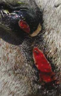
- Skin lesions are ulcerated or raised, progressively enlarging granulomatous masses that bleed easily. Yellow-white gritty plaques are apparent when the lesions are explored or sectioned
- The classical lesion of habronemiasis is an ulcerated or raised granuloma at the medial canthus or on a line from the medial canthus that overlies the course of the nasolacrimal duct. The eyelids may have either ulcerative, granulomatous, or combination lesions, which may originate from either the skin of the eyelids or the palpebral conjunctiva
- Lesions of the conjunctiva are particularly dangerous since the same gritty, calcareous plaques observed in skin lesions occur in ocular adnexal lesions and constitute abrasive foreign bodies that frequently injure the cornea
Diagnosis
Diagnosis is usually based on a combination of environmental factors (season, location) and the characteristic appearance. However, washings taken from the ulcerated area with warm saline under mild pressure and collected into a Petri dish usually reveal the larvae in considerable numbers.
- It is recognised easily by its appearance, i.e. a granulomatous mass with yellow, caseous, gritty contents
- The presence of these plaques helps to distinguish these lesions from fibroplastic sarcoids and exuberant granulation tissue
- If unsure, biopsy to confirm and to rule out neoplasia
Treatment and control
- Ivermectin applied topically or in an oral formulation, e.g. solution such as 0.05% ivermectin in saline
- Systemic therapy with ivermectin at the recommended dose given at three to six week intervals, according to the severity of the disease, kills the larvae and produces rapid resolution of lesions
- The conjunctival form requires more careful treatment using a weak ivermectin solution in balanced, artificial tear solution.
- Topical corticosteroid drops may be needed if there is a severe reaction to the dying larvae
- In conjunction with ivermectin, other therapies, including surgical de-bulking of lesions, debridement of obvious calcareous nodules or plaques, wound dressing and bandaging (to deter granulation tissue when lesions occur over the lower limbs or points of motion) are necessary
- Treatment must also be aimed at decreasing inflammatory reactions. Topical application of a combination of dimethyl sulphoxide and dexamethasone suspended in nitrofuracin ointment have been found to be helpful adjuncts in reducing granulomatous lesions (Rebhun, 1996)
- Fly repellents to control fly strike should also be employed
Literature Search
Use these links to find recent scientific publications via CAB Abstracts (log in required unless accessing from a subscribing organisation).
Habronemiasis in donkeys related publications
References
- Grove, V. (2008) Conditions of the eye In Svendsen, E.D., Duncan, J. and Hadrill, D. (2008) The Professional Handbook of the Donkey, 4th edition, Whittet Books, Chapter 11
- Knottenbelt, D. (2008) Skin disorders In Svendsen, E.D., Duncan, J. and Hadrill, D. (2008) The Professional Handbook of the Donkey, 4th edition, Whittet Books, Chapter 8
- Trawford, A. and Getachew, M. (2008) Parasites In Svendsen, E.D., Duncan, J. and Hadrill, D. (2008) The Professional Handbook of the Donkey, 4th edition, Whittet Books, Chapter 6
- Getachew, M. (1999). ‘Epidemiological study on the health and welfare of Ethiopian donkeys, with particular reference to parasitic diseases’. MVM thesis. Faculty of Veterinary Medicine, University of Glasgow.
- Pandey, V.S., Khallaayoune, K., Ouhelli, H., and Dakkak, A. (1994). ‘Parasites of donkeys in Africa’. M. Bakkoury and R.A. Prentis (eds). ‘Working equines’. Proceedings of 2nd International Colloquium, 22-24 April 1994. Rabat, Morocco. pp 35-44
- Rebhun, W.C. (1996). ‘Observations on habronemiasis in horses’. Equine Veterinary Education 8(4). pp 188-191.
|
|
This section was sponsored and content provided by THE DONKEY SANCTUARY |
|---|
