Therapeutic Techniques for the eye - Donkey
Swab technique
All cases of ulcerative keratitis should be swabbed and the antibiotics selected on the basis of results. Bacterial and fungal cultures should be made from the swabs.
- A moistened swab is recommended to sample an ulcerated cornea. The commercially available swabs contain transport medium that can be used to pre-moisten
- It is important to take the sample from the lesion itself, avoiding the eyelids
- Recurrent or refractive conjunctivitis should be swabbed, ensuring that any medication has been ceased for one to two days prior to sampling
- The need for diagnostic tests should be considered before applying any solutions or stains, as they usually contain preservatives, which will affect culture for bacteria and fungus
- None of the regional nerve blocks reduce corneal sensitivity, so topical anaesthesia is necessary in these cases, but not usually for margins or conjunctiva. If sending the samples to an external laboratory, attention should be drawn to this
- If a keratomycosis is suspected, it is best to discuss the required samples with the laboratory, as a deep corneal scrape may be recommended and the sample must be processed correctly. Obviously, if fungal elements are found, a poorer prognosis can be given and antimycotic treatment can be started promptly
- External laboratories are usually used, but it can be useful to perform a Gram’s and Giemsa stain in-house, thus allowing a rapid decision to be made
Biopsy
Under topical or local anaesthesia, suspicious lesions can be biopsied using fine needle aspiration or surgical excision.
Lavage systems

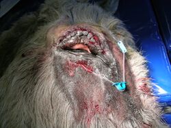
Subpalpebral lavage systems are currently preferred to the nasolacrimal cannulae. One advantage is that they allow the passage of ocular debris to flow in its natural direction. The other advantages are their ease of placement, use and maintenance. They are tolerated well by patients, and can be kept in place happily for more than three weeks. Only 0.2 ml of solution are needed, followed by 0.5 ml of air to clear the system. The flushing of air is an essential part of maintaining the system but the animal may object to the sensation.
Nasolacrimal cannulas require about 1 ml of solution followed by 1 ml of air, as the tube is much wider and longer.
Each system has full instructions on the packaging for placement. The tubing can be secured with ties, glue or sutures. If the mane of the donkey is long enough, then ties can be used but this is often not the case. Sutures rarely fail but take longer to apply and require a bleb of local anaesthetic prior to insertion.
If the donkey is stabled with a companion to which it is bonded, this can shorten the life of the system, unless loose ends are firmly secured and hidden. A donkey that discovers the fun in chewing the cap off can prove quite expensive!
‘Vetwrap’ bandages are useful in retaining the tubing and cap. If the tubing comes loose and the footplate dangles under the eyelid, it causes severe trauma to the cornea, producing a corneal ulcer or worse if not noticed early. The tubing should neither be slack nor pulled tight.
As with all ocular cases, food and hay should be fed from the floor to minimise contamination of the eye. The system should, therefore, be checked in relation to the full range of head extension.
The system must be inspected daily and kept well maintained, preferably by a trained person. Hospitalisation is ideal, but if the owner is competent then it can save disruption to the animal if it is treated in its normal environment.
As the injection port is secured behind the poll, and as far back as the withers, the systems are ideal for head-shy or fractious animals. The medication should be injected slowly so as not to cause alarm or trauma. All treatments should be flushed through with air, leaving a five-minute interval between injections.
The main complication of subpalpebral lavage systems is the development of eyelid abscesses if injected material enters the surrounding tissues. This can cause considerable swelling and demands the immediate removal of the system. Treatment with systemic antibiotics, NSAIDs and gentle antiseptic bathing will normally result in resolution within days.
Subconjunctival injection
In the case of serious infections within the eye, such as a stromal abscess or penetrating foreign body, subconjunctival injections of antibiotic are commonly used to deliver high concentrations of antibiotic through local uptake into the bloodstream and slow release into the tissues around the eye. Other solutions that can also be used are antifungals, atropine and corticosteroids. The technique is best performed using sedation and local anaesthesia due to the potential risks of misdirecting the needle. Use a 23 gauge 16 mm needle. The maximum volume of injection is 1 ml and depot preparations should be avoided.
Literature Search
Use these links to find recent scientific publications via CAB Abstracts (log in required unless accessing from a subscribing organisation).
Ocular Therapeutics in donkeys or horses publications
References
- Grove, V. (2008) Conditions of the eye In Svendsen, E.D., Duncan, J. and Hadrill, D. (2008) The Professional Handbook of the Donkey, 4th edition, Whittet Books, Chapter 11
|
|
This section was sponsored and content provided by THE DONKEY SANCTUARY |
|---|
