Difference between revisions of "Hard Palate"
Jump to navigation
Jump to search
(→Links) |
|||
| (9 intermediate revisions by 2 users not shown) | |||
| Line 1: | Line 1: | ||
| − | |||
==Overview== | ==Overview== | ||
| Line 7: | Line 6: | ||
==Structure and Function== | ==Structure and Function== | ||
| − | The hard palate is the bony shelf of the palatine processes of the [[Skull and Facial Muscles - Anatomy & Physiology#Incisive Bone (os incisivium)|incisive]], [[Skull and Facial Muscles - Anatomy & Physiology#Maxilla|maxillary]] and [[Skull and Facial Muscles - Anatomy & Physiology#Paltine Bone (os palatinium)|palatine bones]]. Failure of the [[Skull and Facial Muscles - Anatomy & Physiology# | + | The hard palate is the bony shelf of the palatine processes of the [[Skull and Facial Muscles - Anatomy & Physiology#Incisive Bone (os incisivium)|incisive]], [[Skull and Facial Muscles - Anatomy & Physiology#Maxilla|maxillary]] and [[Skull and Facial Muscles - Anatomy & Physiology#Paltine Bone (os palatinium)|palatine bones]]. Failure of the [[Skull and Facial Muscles - Anatomy & Physiology#Paltine Bone (os palatinium)|palatine bones]] to fuse results in [[Cleft Palate|cleft palate]]. |
There are 6-8 fixed transverse ridges to direct food caudally. The hard palate is flat and has '''incisive papilla''' (small median swelling) behind the incisive [[:Category:Teeth - Anatomy & Physiology|teeth]] and smaller '''papillae ducts''' branching to the [[Nasal Cavity - Anatomy & Physiology|nasal cavity]] and vomeronasal organ. | There are 6-8 fixed transverse ridges to direct food caudally. The hard palate is flat and has '''incisive papilla''' (small median swelling) behind the incisive [[:Category:Teeth - Anatomy & Physiology|teeth]] and smaller '''papillae ducts''' branching to the [[Nasal Cavity - Anatomy & Physiology|nasal cavity]] and vomeronasal organ. | ||
| Line 13: | Line 12: | ||
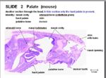
[[Image:Hard Palate Histology.jpg|thumb|right|150px|Hard Palate (Mouse) - Copyright RVC 2008]] | [[Image:Hard Palate Histology.jpg|thumb|right|150px|Hard Palate (Mouse) - Copyright RVC 2008]] | ||
| − | + | *Thick mucosa | |
| + | |||
| + | *keratinised stratified squamous epithelium | ||
==Species Differences== | ==Species Differences== | ||
| Line 25: | Line 26: | ||
==Links== | ==Links== | ||
| − | Click here for the [[Cleft Palate|Pathology of Cleft Palate]] | + | Click here for the [[Cleft Palate|Pathology of Cleft Palate]] and hard palate [[Hard Palate - Histology|histology]]. |
| − | |||
| − | |||
[[Category:Oral Cavity - Anatomy & Physiology]] | [[Category:Oral Cavity - Anatomy & Physiology]] | ||
| + | [[Category:To Do - AimeeHicks]][[Category:To Do - AP Review]] | ||
Revision as of 16:59, 10 December 2010
Overview
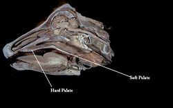
The hard palate (palatum durum) forms the rostral roof of the oral cavity. It merges caudally with the soft palate where a connective tissue aponeurosis replaces the bone.
Structure and Function
The hard palate is the bony shelf of the palatine processes of the incisive, maxillary and palatine bones. Failure of the palatine bones to fuse results in cleft palate. There are 6-8 fixed transverse ridges to direct food caudally. The hard palate is flat and has incisive papilla (small median swelling) behind the incisive teeth and smaller papillae ducts branching to the nasal cavity and vomeronasal organ.
Histology
- Thick mucosa
- keratinised stratified squamous epithelium
Species Differences
Herbivores
Herbivores have a more heavily keratinised hard palate.
Feline
Felines have short a hard palate.
Links
Click here for the Pathology of Cleft Palate and hard palate histology.

