Difference between revisions of "Equine Encephalitis Virus"
| Line 1: | Line 1: | ||
| − | {{ | + | {{review}} |
| + | |||
Also known as: '''''EEV — Alphavirus — Eastern Equine Encephalitis Virus, EEE — Western Equine Encephalitis Virus, WEE — Venezuelan Equine Encephalitis Virus, VEE | Also known as: '''''EEV — Alphavirus — Eastern Equine Encephalitis Virus, EEE — Western Equine Encephalitis Virus, WEE — Venezuelan Equine Encephalitis Virus, VEE | ||
| Line 43: | Line 44: | ||
*Virus isolation is the most definitive diagnostic method for EEE or WEE.<ref name="again">''Manual of Diagnostic Tests and Vaccines for Terrestrial Animals'' found at http://www.oie.int/eng/normes/mmanual/A_00081.htm, accessed July 2010.</ref>. Brain is preferred, but virus has also been isolated from the liver and spleen.<ref name="again">''Manual of Diagnostic Tests and Vaccines for Terrestrial Animals'' found at http://www.oie.int/eng/normes/mmanual/A_00081.htm, accessed July 2010.</ref>. Samples of these tissues should be taken in duplicate, one set for virus isolation and the other placed in formalin for histopathology.<ref name="again">''Manual of Diagnostic Tests and Vaccines for Terrestrial Animals'' found at http://www.oie.int/eng/normes/mmanual/A_00081.htm, accessed July 2010.</ref>. Viral isolation specimens should be sent frozen unless they can be received refrigerated within 48 hours of sampling.<ref name="again">''Manual of Diagnostic Tests and Vaccines for Terrestrial Animals'' found at http://www.oie.int/eng/normes/mmanual/A_00081.htm, accessed July 2010.</ref>. Unless clinical signs persist for more than 5days prior to death, EEE virus is frequently isolated from equine brain tissue. WEE virus, however, is rarely isolated from tissues of infected horses. Newborn mice, chicken embryos and a number of cell culture systems can be used for virus isolation.<ref name="again">''Manual of Diagnostic Tests and Vaccines for Terrestrial Animals'' found at http://www.oie.int/eng/normes/mmanual/A_00081.htm, accessed July 2010.</ref>. Virus may also be isolated from cerebrospinal fluid (CSF) of acutely infected horses.<ref name="duplicate">Merck & Co (2008) The Merck Veterinary Manual (Eighth Edition), Merial found at http://www.merckvetmanual.com/mvm/index.jsp?cfile=htm/bc/100900.htm&word=Equine%2cencephalitis, accessed July 2010</ref> | *Virus isolation is the most definitive diagnostic method for EEE or WEE.<ref name="again">''Manual of Diagnostic Tests and Vaccines for Terrestrial Animals'' found at http://www.oie.int/eng/normes/mmanual/A_00081.htm, accessed July 2010.</ref>. Brain is preferred, but virus has also been isolated from the liver and spleen.<ref name="again">''Manual of Diagnostic Tests and Vaccines for Terrestrial Animals'' found at http://www.oie.int/eng/normes/mmanual/A_00081.htm, accessed July 2010.</ref>. Samples of these tissues should be taken in duplicate, one set for virus isolation and the other placed in formalin for histopathology.<ref name="again">''Manual of Diagnostic Tests and Vaccines for Terrestrial Animals'' found at http://www.oie.int/eng/normes/mmanual/A_00081.htm, accessed July 2010.</ref>. Viral isolation specimens should be sent frozen unless they can be received refrigerated within 48 hours of sampling.<ref name="again">''Manual of Diagnostic Tests and Vaccines for Terrestrial Animals'' found at http://www.oie.int/eng/normes/mmanual/A_00081.htm, accessed July 2010.</ref>. Unless clinical signs persist for more than 5days prior to death, EEE virus is frequently isolated from equine brain tissue. WEE virus, however, is rarely isolated from tissues of infected horses. Newborn mice, chicken embryos and a number of cell culture systems can be used for virus isolation.<ref name="again">''Manual of Diagnostic Tests and Vaccines for Terrestrial Animals'' found at http://www.oie.int/eng/normes/mmanual/A_00081.htm, accessed July 2010.</ref>. Virus may also be isolated from cerebrospinal fluid (CSF) of acutely infected horses.<ref name="duplicate">Merck & Co (2008) The Merck Veterinary Manual (Eighth Edition), Merial found at http://www.merckvetmanual.com/mvm/index.jsp?cfile=htm/bc/100900.htm&word=Equine%2cencephalitis, accessed July 2010</ref> | ||
| − | + | ==Literature Search== | |
| − | | | + | [[File:CABI logo.jpg|left|90px]] |
| + | |||
| + | |||
| + | Use these links to find recent scientific publications via CAB Abstracts (log in required unless accessing from a subscribing organisation). | ||
| + | <br><br><br> | ||
| + | [http://www.cabdirect.org/search.html?q=title%3A%28%22Equine+Encephalitis+Virus%22%29+AND+od%3A%28horses%29 Equine encephalitis virus in horses] | ||
[http://www.cabdirect.org/search.html?rowId=1&options1=AND&q1=%22Equine+Encephalitis+Virus%22&occuring1=title&rowId=2&options2=NOT&q2=horses&occuring2=od&rowId=3&options3=AND&q3=&occuring3=freetext&x=42&y=9&publishedstart=yyyy&publishedend=yyyy&calendarInput=yyyy-mm-dd&la=any&it=any&show=all Equine encephalitis virus in other species, including laboratory animals] | [http://www.cabdirect.org/search.html?rowId=1&options1=AND&q1=%22Equine+Encephalitis+Virus%22&occuring1=title&rowId=2&options2=NOT&q2=horses&occuring2=od&rowId=3&options3=AND&q3=&occuring3=freetext&x=42&y=9&publishedstart=yyyy&publishedend=yyyy&calendarInput=yyyy-mm-dd&la=any&it=any&show=all Equine encephalitis virus in other species, including laboratory animals] | ||
| − | |||
==References== | ==References== | ||
| Line 53: | Line 58: | ||
<references/> | <references/> | ||
| − | |||
| − | |||
| − | |||
| − | |||
[[Category:Togaviridae]] | [[Category:Togaviridae]] | ||
Revision as of 13:22, 7 January 2011
| This article has been peer reviewed but is awaiting expert review. If you would like to help with this, please see more information about expert reviewing. |
Also known as: EEV — Alphavirus — Eastern Equine Encephalitis Virus, EEE — Western Equine Encephalitis Virus, WEE — Venezuelan Equine Encephalitis Virus, VEE
Introduction
Family Togaviridae
Genus Alphavirus
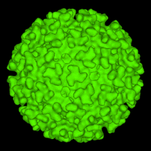
Single-stranded, linear, positive-sense RNA viruses, 60-70nm in diameter.[1]
Important Serotypes
Several alphavirus strains have been isolated during equine and human epidemics of encephalitis in the Western Hemisphere. These epidemics have most often been attributed to:
- Eastern Equine Encephalitis virus (EEV)
- Western EEV
- Venezuelan EEV.[1]
Eastern EEV has North and South American antigenic variants.[2] Western EEV is a recombinant between an Eastern EEV-like virus and a Sindbis-like virus.[3] Western EEV also has two antigenic subtypes - WEE and Highlands J viruses.[1] Considerable overlap exists between the various strains in terms of their geography, and potentially also in their antigenic properties and biological behaviour.[2] Of the 6 subtypes of Venezuelan EEV (I-VI), significant outbreaks of equine encephalitis in the Western Hemisphere over the last two decades have been caused by IAB, IC and IE. Variant ID from Central America and variant IF from Brazil are considered endemic and typically demonstrate low pathogenicity for horses. These features are also typical of subtype II (Everglades) virus in Florida and types II, IV, V and VI viruses.[1]
Diseases
Arboviruses are the most common cause of equine encephalitis.[4]
Distribution
Virus distribution is largely dictated by the distribution of appropriate vectors. Thus Eastern EEV can be found in a region extending from eastern Canada at its northern extremity, down throughout the Caribbean and in parts of Central and South America.[1]. The virus has also been identifed in the Philippines[5] and may be present in Europe.[6] Over the last two decades, Western EEV has rarely caused disease in the western United States, although it is found here in avian reservoir hosts.[1] As well as producing disease outbreaks in Mexico, Venezuela and Colombia, Venezuelan EEV has recently been recognized in Peru, Brazil, French Guiana and Trinidad.[1].
Reservoirs
Togaviridae survive by asymptomatically infecting sylvatic hosts such as birds, small mammals, reptiles and amphibians.[1] Overwintering occurs in these wild populations.
Vectors
During a blood meal, mosquito vectors transmit viral particles between wild animal hosts. Viruses that are able to penetrate the mosquito's gut can pass via the haemolymph to the oral glands. Here, viral replication is followed by the shedding of viral particles into the saliva and other oral secretions. Viral multiplication may not be necessary for transmission if the original blood meal contained sufficient numbers of viral particles.[1]. It is thought that the mosquito remains permanently infected.[7] The most significant disease vectors for each viral serotype are:
- Eastern EEV: Aedes spp.
- Western EEV: Culex tarsalis
- Venezuelan EEV: Culex melanconium, Aedes spp., Phosphora spp.[1].
Culiseta melanura is another vector for Eastern EEV. It feeds mostly on swamp birds, completing an enzootic cycle of viral transmission. C.melanura is thus an inhabitant of freshwater swamps and is not usually found in areas densely populated by equids.[8] Epizootics and epidemics of Eastern EEV disease are propagated by Aedes spp. Western EEV persists in an enzootic cycle with passerine birds, transmitted by C.tarsalis.[1]. Other vectors or overwintering hosts for this serotype may include Dermacentor andersoni ticks[9], Triatoma sanguisuga (the assassin bug)[10], and the cliff swallow bug (Oeciacus vicarius).[11] Epidemic strains of Venezuelan EEV have infected mosquito species from several genera and this viral serotype may also be transmitted by ticks.[1].
Virus Identification
Viruses can be identified by complement fixation, immunofluorescence, PCR, ELISA, or virus isolation.
- The complement fixation test can be used to identify Eastern or Western EEV in infected mouse or chicken brains, cell culture fluid or amniotic-allantoic fluid.[12]
- Virus may be identified in brain tissue or cell culture using direct immunofluorescent staining.[12]
- EEE and WEE viral RNA in mosquitoes and equine tissues may be detected by reverse-transcription PCR.[13]
- ELISA can be used to detect virus in brain tissue. An antigen-capture ELISA, developed for EEE surveillance in mosquitoes, can be used where virus isolation and PCR facilities are unavailable. [14]
- Virus isolation is the most definitive diagnostic method for EEE or WEE.[12]. Brain is preferred, but virus has also been isolated from the liver and spleen.[12]. Samples of these tissues should be taken in duplicate, one set for virus isolation and the other placed in formalin for histopathology.[12]. Viral isolation specimens should be sent frozen unless they can be received refrigerated within 48 hours of sampling.[12]. Unless clinical signs persist for more than 5days prior to death, EEE virus is frequently isolated from equine brain tissue. WEE virus, however, is rarely isolated from tissues of infected horses. Newborn mice, chicken embryos and a number of cell culture systems can be used for virus isolation.[12]. Virus may also be isolated from cerebrospinal fluid (CSF) of acutely infected horses.[4]
Literature Search
Use these links to find recent scientific publications via CAB Abstracts (log in required unless accessing from a subscribing organisation).
Equine encephalitis virus in horses
Equine encephalitis virus in other species, including laboratory animals
References
- ↑ 1.00 1.01 1.02 1.03 1.04 1.05 1.06 1.07 1.08 1.09 1.10 1.11 Bertone, J.J (2010) Viral Encephalitis in Reed, S.M, Bayly, W.M. and Sellon, D.C (2010) Equine Internal Medicine (Third Edition), Saunders, Chapter 12
- ↑ 2.0 2.1 Casal, J (1964) Antigenic variants of equine encephalitis virus, J Exp Med 119:547-565. In: Bertone, J.J (2010) Viral Encephalitis in Reed, S.M, Bayly, W.M. and Sellon, D.C (2010) Equine Internal Medicine (Third Edition), Saunders, Chapter 12.
- ↑ Hahn, C.S, Lustig, S, Strauss, E.G, Strauss, J.H (1988) Western equine encephalitis virus is a recombinant virus, Proc Natl Acad Sci USA 85(16):5997-6001. In: Bertone, J.J (2010) Viral Encephalitis in Reed, S.M, Bayly, W.M. and Sellon, D.C (2010) Equine Internal Medicine (Third Edition), Saunders, Chapter 12.
- ↑ 4.0 4.1 Merck & Co (2008) The Merck Veterinary Manual (Eighth Edition), Merial found at http://www.merckvetmanual.com/mvm/index.jsp?cfile=htm/bc/100900.htm&word=Equine%2cencephalitis, accessed July 2010
- ↑ Livesay, H.R (1949) Isolation of eastern equine encephalitis virus from naturally infected monkey (Macacus philippensis), J Infect Dis 84:306-309. In: Bertone, J.J (2010) Viral Encephalitis in Reed, S.M, Bayly, W.M. and Sellon, D.C (2010) Equine Internal Medicine (Third Edition), Saunders, Chapter 12
- ↑ Gibbs, E.P.J (1976) Equine viral encephalitis, Equine Vet J, 8:66-71. In: Bertone, J.J (2010) Viral Encephalitis in Reed, S.M, Bayly, W.M. and Sellon, D.C (2010) Equine Internal Medicine (Third Edition), Saunders, Chapter 12
- ↑ Crans, W.J, McNeilly, J, Schulze, T.L, Main, A (1986)Isolation of eastern equine encephalitis virus from Aedes solicitans during an epizootic in Southern New Jersey, J Am Mosq Control Assoc 2:68-72. In: Bertone, J.J (2010) Viral Encephalitis in Reed, S.M, Bayly, W.M. and Sellon, D.C (2010) Equine Internal Medicine (Third Edition), Saunders, Chapter 12
- ↑ Hoff, G.L, Bigler, W.J, Buff, E.E, Beck, E (1978) Occurrence and distribution of western equine encephalomyelitis in Florida, J Am Vet Med Assoc, 172:351-352. In: Bertone, J.J (2010) Viral Encephalitis in Reed, S.M, Bayly, W.M. and Sellon, D.C (2010) Equine Internal Medicine (Third Edition), Saunders, Chapter 12
- ↑ Syverton, J.T, Berry, G.P (1937) The tick as a vector for the virus disease equine encephalomyelitis, J Bacteriol, 33:60. In: Bertone, J.J (2010) Viral Encephalitis in Reed, S.M, Bayly, W.M. and Sellon, D.C (2010) Equine Internal Medicine (Third Edition), Saunders, Chapter 12
- ↑ Kitselman, C.H, Grundman, A.W (1940) Equine encephalomyelitis virus isolated from naturally infected Triatoma sanguisuga, Kans Agric Exp Station Tech Bull, 50:15. In: Bertone, J.J (2010) Viral Encephalitis in Reed, S.M, Bayly, W.M. and Sellon, D.C (2010) Equine Internal Medicine (Third Edition), Saunders, Chapter 12
- ↑ Hayes, R.O, Francy, D.B, Lazuick, J.S (1977) Role of the cliff swallow bug (Oeciacus vicarius) in the natural cycle of a Western equine encephalitis-related alphavirus, J Entomol, 14:257-262. In: Bertone, J.J (2010) Viral Encephalitis in Reed, S.M, Bayly, W.M. and Sellon, D.C (2010) Equine Internal Medicine (Third Edition), Saunders, Chapter 12
- ↑ 12.0 12.1 12.2 12.3 12.4 12.5 12.6 Manual of Diagnostic Tests and Vaccines for Terrestrial Animals found at http://www.oie.int/eng/normes/mmanual/A_00081.htm, accessed July 2010.
- ↑ Vodkin, M.H, McLaughlin, G.L, Day, J.F, Shope, R.E, Novak, R.J (1993) A rapid diagnostic assay for eastern equine encephalomyelitis viral-RNA, Am J Trop Med Hyg, 49, 772-776. In: Manual of Diagnostic Tests and Vaccines for Terrestrial Animals found at http://www.oie.int/eng/normes/mmanual/A_00081.htm, accessed July 2010.
- ↑ Brown, T.M, Mitchell, C.J, Nasci, R.S, Smith, G.C. and Roehrig, J.T. (2001). Detection of eastern equine encephalitis virus in infected mosquitoes using a monoclonal antibody-based antigen-capture enzyme-linked immunosorbent assay, Am J Trop Med Hyg, 65, 208-213. In: Manual of Diagnostic Tests and Vaccines for Terrestrial Animals found at http://www.oie.int/eng/normes/mmanual/A_00081.htm, accessed July 2010.

