Difference between revisions of "Equine Internal Medicine Q&A 05"
| (2 intermediate revisions by the same user not shown) | |||
| Line 18: | Line 18: | ||
In the horse the deposits are most severe within the glomerular capillary walls. <br> | In the horse the deposits are most severe within the glomerular capillary walls. <br> | ||
These deposits cause activation of soluble and cellular mediators within the kidney, causing protein loss in the urine, decreased glomerular filtration and renal failure. | These deposits cause activation of soluble and cellular mediators within the kidney, causing protein loss in the urine, decreased glomerular filtration and renal failure. | ||
| − | |l2=Glomerulonephritis | + | |l2=Glomerulonephritis#Aetiology |
| + | |||
</FlashCard> | </FlashCard> | ||
Revision as of 13:57, 22 August 2011
| This question was provided by Manson Publishing as part of the OVAL Project. See more Equine Internal Medicine questions |
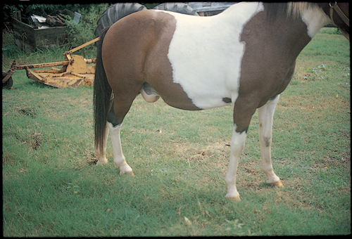
Shown is an eight-year-old pony gelding with weight loss (approximately 45kg), inappetence and non-painful pitting oedema of the ventral abdomen. The heart sounds normal, the heart rate is 40bpm and the jugular veins are not distended. The pony is azotaemic (creatinine 194.5␣mol/l; 2.2mg/dl) and the urinalysis reveals specific gravity of 1.014 and proteinuria (urine protein:creatinine ratio 3:1). The PCV of the pony is 35% and the plasma protein is 45g/l.
| Question | Answer | Article | |
| What is the most likely diagnosis in this pony? | The most likely diagnosis is glomerulonephritis with renal failure. |
Link to Article | |
| What is the presumed pathological mechanism of the disease? | The presumed pathological mechanism of glomerulonephritis is persistent immune complex deposition or in situ formation in the glomerulus. In the horse the deposits are most severe within the glomerular capillary walls. |
[[Glomerulonephritis#Aetiology |Link to Article]] | |
