Difference between revisions of "Veterinary Dentistry Q&A 01"
(No difference)
| |
Latest revision as of 22:49, 12 October 2011
| This question was provided by Manson Publishing as part of the OVAL Project. See more Veterinary Dentistry Q&A. |
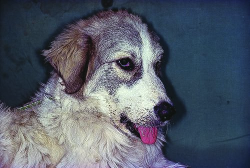
This dog was presented with an acute inability to close the mouth but no lateral deviation of the mandible.
| Question | Answer | Article | |
| What is your tentative clinical diagnosis? | Bilateral traumatic luxation of the temporomandibular joint. |
Link to Article | |
| How can this diagnosis be confirmed? | The diagnosis is confirmed by radiography. Two views are currently in use: the dorsoventral closed-mouth skull radiograph and the closed-mouth lateral oblique view (15–20°, nose tilted up). Unilateral or bilateral luxation is radiologically evidenced by the fact that the condyloid process is not located within the mandibular fossa. Capsular osteophyte formation is evidence of a long-standing or recurrent luxation. Usually, the condyloid process displaces rostrodorsally. If unilateral, the animal is presented with a typical lateral deviation to the side opposite the luxated joint. |
Link to Article | |
| Presuming that the tentative diagnosis is confirmed, what is the treatment? | Reduction is accomplished under general anesthesia by forcing the condyle ventrally. This can be done by inserting a fulcrum (e.g. pencil, syringe, dowel – depending on patient size) in between the molar teeth and gently forcing the mouth closed; this in turn levers the condyloid process in a ventrocaudal direction back into the condyloid fossa. Aftercare may include the use of a tape muzzle. Recurrent and chronic luxations can be treated by condylectomy. |
Link to Article | |
