Difference between revisions of "Veterinary Dentistry Q&A 11"
Ggaitskell (talk | contribs) |
|||
| (4 intermediate revisions by 2 users not shown) | |||
| Line 2: | Line 2: | ||
|book = Veterinary Dentistry Q&A}} | |book = Veterinary Dentistry Q&A}} | ||
| − | [[ | + | [[File:Vet Dentistry 11.jpg|centre|500px]] |
<br /> | <br /> | ||
| Line 20: | Line 20: | ||
Where it represents pathology, it is an extension of a chronic pulpitis or an inflammatory reaction due to pulpal necrosis. | Where it represents pathology, it is an extension of a chronic pulpitis or an inflammatory reaction due to pulpal necrosis. | ||
| − | |l1= | + | |l1= |
|q2=How would you determine whether the described lesions are associated with disease? | |q2=How would you determine whether the described lesions are associated with disease? | ||
|a2= | |a2= | ||
| − | The infrabony pocket can | + | The infrabony pocket can by confirmed by periodontal probing and is associated with the presence of periodontitis. The fact that the periapical radiolucency appears to be an extension of the root shape, rather than a round ballooning lesion, is suggestive |
| + | for normal anatomy. | ||
Comparison should always be made with other teeth of the same type in the same animal. A contralateral radiograph is indicated, particularly if the tooth seems healthy, e.g. no sign of crown fracture or pulpal involvement, on clinical examination. | Comparison should always be made with other teeth of the same type in the same animal. A contralateral radiograph is indicated, particularly if the tooth seems healthy, e.g. no sign of crown fracture or pulpal involvement, on clinical examination. | ||
| − | |l2= | + | |l2= |
</FlashCard> | </FlashCard> | ||
Revision as of 22:52, 12 October 2011
| This question was provided by Manson Publishing as part of the OVAL Project. See more Veterinary Dentistry Q&A. |
| Question | Answer | Article | |
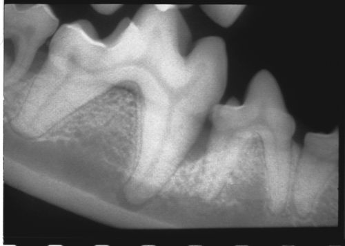
| Describe the radiologic findings on this radiograph. | A deep infrabony pocket is evident on the mesial aspect of the mesial root of the mandibular first molar, also involving the distal root of the fourth premolar. In addition, there is a distinct periapical radiolucent area associated with the same root. This lesion may be normal anatomy or represent pathology. Where it represents normal anatomy, it is a radiographic artifact due to the root apex dipping into the mandibular canal or lying in close proximity to the canal. Where it represents pathology, it is an extension of a chronic pulpitis or an inflammatory reaction due to pulpal necrosis. |
[[|Link to Article]] | |
| How would you determine whether the described lesions are associated with disease? | The infrabony pocket can by confirmed by periodontal probing and is associated with the presence of periodontitis. The fact that the periapical radiolucency appears to be an extension of the root shape, rather than a round ballooning lesion, is suggestive for normal anatomy. Comparison should always be made with other teeth of the same type in the same animal. A contralateral radiograph is indicated, particularly if the tooth seems healthy, e.g. no sign of crown fracture or pulpal involvement, on clinical examination. |
[[|Link to Article]] | |
