Difference between revisions of "Veterinary Dentistry Q&A 04"
Ggaitskell (talk | contribs) |
Ggaitskell (talk | contribs) |
(No difference)
| |
Latest revision as of 07:49, 14 October 2011
| This question was provided by Manson Publishing as part of the OVAL Project. See more Veterinary Dentistry Q&A. |
Discolorations of teeth can be classified as generalized, local, or pseudodiscolorations.
| Question | Answer | Article | |
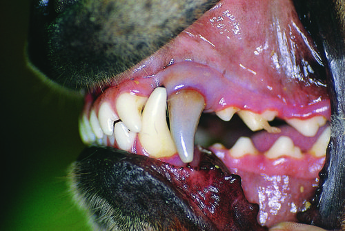
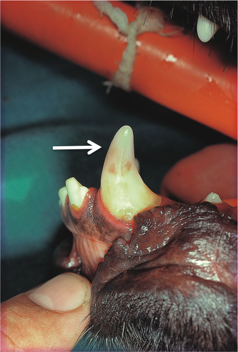
| Which type can be seen here, and what are the possible causes? | The list below summarizes the causes of dental discoloration. The case in question is a local discoloration, probably of endodontic–traumatic origin. Hemorrhage or necrosis of the pulp causes lysis of the erythrocytes. The hemoglobin breaks down into pigments which penetrate the dentinal tubules and are responsible for the different discolorations. The color of the crown may vary from pink–red to blue–gray or dark gray. In the case of a minor hemorrhage, the pulp may survive, blood pigments may be resorbed and the discoloration may be transient. Another possible cause of crown discoloration is acute pulpitis of hematogenous origin. A localized pink–red area on the crown may be indicative of vital pulp with internal resorption, the vascular resorbing tissue being visible through the enamel (‘pink spot’ – 36b, arrowed). Generalized discolorations:
Local discolorations:
Pseudodiscolorations:
|
Link to Article | |
| Is treatment necessary and, if performed, will the discoloration disappear? | Radiographic examination and pulp-vitality testing (if available) are indicated and endodontic treatment is most often the treatment of choice. It is important to note that discoloration may persist following endodontic treatment. |
Link to Article | |

