Difference between revisions of "Tritrichomonas foetus"
m |
|||
| (2 intermediate revisions by the same user not shown) | |||
| Line 1: | Line 1: | ||
| − | |||
{{Taxobox | {{Taxobox | ||
|kingdom =Protista | |kingdom =Protista | ||
| Line 24: | Line 23: | ||
The protozoa resides on the surface and in the lumen of the female reproductive tract and in the crypt of penile epithelial cells. Transmission is achieved from infected individuals during mating and direct contact. | The protozoa resides on the surface and in the lumen of the female reproductive tract and in the crypt of penile epithelial cells. Transmission is achieved from infected individuals during mating and direct contact. | ||
| − | ''T. foetus'' has no known cyst stage, although carrier cows occur very rarely and the pathogenesis of the carrier state is unknown. Therefore, trophozoites replicate simply by binary fission. It is thought that ''T. foetus'' feeds from host lipids and fatty acids <ref>Beach DH, Holz GGJr, Singh BN, Lindmark DG, 1991. '''Phospholipid metabolism of cultured ''Trichomonas vaginalis'' and | + | ''T. foetus'' has no known cyst stage, although carrier cows occur very rarely and the pathogenesis of the carrier state is unknown. Therefore, trophozoites replicate simply by binary fission. It is thought that ''T. foetus'' feeds from host lipids and fatty acids <ref>Beach DH, Holz GGJr, Singh BN, Lindmark DG, 1991. '''Phospholipid metabolism of cultured ''Trichomonas vaginalis'' and Tritrichomonas foetus'''. ''Molecular and Biochemical Parasitology'', 44(1:97-108;19)</ref> |
''T. foetus'' is sometimes isolated from foetal gut and lung but this is unlikely to be due to invasion and more likely due to swallowing or inhalation of amniotic fluid in utero. | ''T. foetus'' is sometimes isolated from foetal gut and lung but this is unlikely to be due to invasion and more likely due to swallowing or inhalation of amniotic fluid in utero. | ||
| Line 66: | Line 65: | ||
{{Nick Lyons | {{Nick Lyons | ||
|date = 16 October 2011}} | |date = 16 October 2011}} | ||
| − | |||
| − | |||
| − | |||
[[Category:Miscellaneous_Protozoa]] | [[Category:Miscellaneous_Protozoa]] | ||
| − | [[Category:CABI Expert Review Completed | + | [[Category:CABI Expert Review Completed]] |
[[Category:Nick Lyons reviewed]] | [[Category:Nick Lyons reviewed]] | ||
Revision as of 21:27, 29 November 2011
| Tritrichomonas foetus | |
|---|---|
| Kingdom | Protista |
| Phylum | Protozoa |
| Family | Trichomonadidae |
| Genus | Tritrichomonas |
| Species | T. foetus and more |
Also known as: Trichomonas foetus — Trichomonas fetus
Introduction
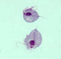
Tritrichomonas foetus is a protozoal pathogen causing disease primarily in cattle and felids. It is identifiable by its pear-like shape, three anterior flagellae (recurrent flagellum) and an undulating membrane on one side.
Lifecycle and Transmission
Cattle
Tritrichomonas foetus is a venereal disease in cattle causing early embryonic death and abortion.
The protozoa resides on the surface and in the lumen of the female reproductive tract and in the crypt of penile epithelial cells. Transmission is achieved from infected individuals during mating and direct contact.
T. foetus has no known cyst stage, although carrier cows occur very rarely and the pathogenesis of the carrier state is unknown. Therefore, trophozoites replicate simply by binary fission. It is thought that T. foetus feeds from host lipids and fatty acids [1]
T. foetus is sometimes isolated from foetal gut and lung but this is unlikely to be due to invasion and more likely due to swallowing or inhalation of amniotic fluid in utero.
Cats
Tritrichomonas foetus is transmitted via the faecal: oral route in cats. Behaviours such as grooming may aid transmission by direct contact. The venereal route is unproven in cats.
Pathogenesis
Cattle
Proteinases secreted by T. foetus degrade substrates such as fibrinogen, albumin and immunoglobulins, thus altering the biological environment and resulting in embryonic death. Pyometra is occasionally observed in females but males show no clinical signs. For more information see Trichomonosis - Cattle.
Cats
T. foetus mainly causes diarrhoea by colonisation of the large intestine. Recovery is slow but longterm prognosis is good. Reports of resistance and success are available for most anthelmintic and antibiotic therapies.
Other trichomonad spp.
Many other trichomonads reside as commensals in the gastrointestinal tract of other species. Two of the most well studied trichomonads are Trichomonas gallinae, which occurs in birds, and Trichomonas vaginalis, which is a common sexually transmitted infection of humans.
Relevant Links: Trichomonosis - Cattle
| Tritrichomonas foetus Learning Resources | |
|---|---|
 Test your knowledge using flashcard type questions |
Protozoa Flashcards Tritrichomonas foetus Flashcards |
References
- ↑ Beach DH, Holz GGJr, Singh BN, Lindmark DG, 1991. Phospholipid metabolism of cultured Trichomonas vaginalis and Tritrichomonas foetus. Molecular and Biochemical Parasitology, 44(1:97-108;19)
Feline Advisory Bureau, Tritrichomonas infection in cats, accessed 02/06/2011 @ http://www.fabcats.org/breeders/infosheets/tritrichomonas/
Wright, I.(2010) Tritrichomonas foetus: Jumping the Species Barrier, UK Vet, Vol 15, 1-3.

|
This article was originally sourced from The Animal Health & Production Compendium (AHPC) published online by CABI during the OVAL Project. The datasheet was accessed on 2 June 2011. |
| This article has been expert reviewed by Nick Lyons MA VetMB CertCHP MRCVS Date reviewed: 16 October 2011 |
