Difference between revisions of "Central Nervous System Neoplasia"
Jump to navigation
Jump to search
(No difference)
| |
Latest revision as of 17:40, 8 November 2012
Neoplasia
- Particularly affects older animals.
- Signs may occur acutely, or be progressive and reflect
- The primary parenchymal damage by the tumour
- Sequelae such as haemorrhage or oedema.
Primary
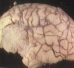
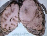
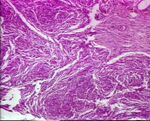
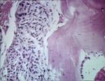
Meningioma
- Meningioma is most frequently seen in cats and dogs, and is the most common primary brain tumour in these species.
- Dolicocephalic dog breeds are predisposed.
- Tumours arise from arachnoid cap cells ion the arachnoid layer of meninges.
- Meningiomas are usually benign, and therefore seldom invade.
- Spread to the lung has, however, been reported.
- The main effects of the tumour is due to its action as a compressive, space-occupying lesion.
- Meningiomas may become mineralised.
| Feature | Dog | Cat |
|---|---|---|
| Lesion Number | Solitary | Multiple |
| Infiltration to Cortical Parenchyma | More infiltrative | Less infiltrative |
| Encapsulation | Poorly encapsulated | Well encapsulated |
| Metastatic Potential | Low | Low |
View images courtesy of Cornell Veterinary Medicine
Treatment
- Chemotherapy
- Radiation therapy
- Surigcal resection.
- Better results in cats (as encapsulated and clearly distinguished from normal brain).
- Survival is 22-27 months following resection.
- Better results in cats (as encapsulated and clearly distinguished from normal brain).
Glioma
- Due to their origin, gliomas are found within the intraaxial neuroxis.
- Brachycephalic breeds are predisposed.
- Glial tumours rise from cells of the brain parenchyma.
- Astrocytes - Astrocytoma
- Oligodendrocytes - Oligodendroglioma
- Ependymal cells - Ependymoma
- Choroid plexus cells - Choroid plexus tumours
Astrocytoma
- The most common of the glial tumors
- Brachycephalic breeds are predisposed.
- E.g. boxer, bulldog.
Gross
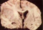
- Astrocytomas are firm, solid tumours.
- Colour tends to be grey-white.
- This may sometimes be mottled with red due to areas of necrosis and haemorrhage.
View images courtesy of Cornell Veterinary Medicine
Oligodendroglioma
- Oligodendroglioma is most commonly found in dogs.
- As for astrocytomas, there is a predilection for brachycephalic breeds.
Gross
- Oligodendrogliomas are soft in texture, and often gelatinous.
- Colour ranges from grey to pink/red.
View images courtesy of Cornell Veterinary Medicine
Ependymoma
- Ependymomas are found in dogs, cats, horses and cattle.
- They occur mainly in the ventricles.
- The lateral ventricle is most often affected.
- The tumours may spread within the ventricular system via the cerebrospinal fluid.
- Growth is generally expansile, but it can be invasive and destructive.
View images courstesy of Cornell Veterinary Medicine
Choroid Plexus Tumours
- Choroid plexus tumours are rare.
- They are mainly found in dogs.
- Choroid plexus tumours are found in areas where the choroid plexus is concentrated, i.e.:
- Lateral ventricle
- Third ventricle
- Fourth ventricle
- There is a particular predilection for the fourth ventricle.
- This association with the ventricular system makes hydrocephalus a common sequelae.
- The tumours may metastasis via the CSF and ventricular system.
- Chroid plexus tumourc contain an increased concentration of blood vessels.
- Contrast administration may therefore aid in their identification.
Treatment of Gliomas
- The usual modes of anti-cancer therapy may be used to tackle gliomas:
- Radiation therapy
- Chemotherapy
- Surgery
- However, surgery is less ideal as the tumours are located within the parenchyma.
PNETs
- PNETs stands for Primitive NeuroEctodermal Tumors.
- These are tumors of primitive germ cell origin.
- They are rare.
Secondary
- May arise from:
- Metastasis
- Numerous tumours of older animals may metastasise to the brain:
- Haemangiosarcoma
- Lymphoma
- Mammary gland carcinomas
- Other carcinomas
- Tumours which metastasise to the lungs may be more likely to metastasise to the brain.
- Incidence is underestimated, as the brain is not routinely examined at necropsy.
- The white-grey matter junction is the most frequently affected area.
- Brainstem and spinal cord metastasis are less common than forebrain metastasis.
- Choroid plexus tumours and ependymomas may metastasise via the CSF.
- Numerous tumours of older animals may metastasise to the brain:
- Extenstion from extraneural sites, e.g.
- Skull
- Nasal cavity
- Signs of extenstion may preced signs of nasal disease.
- Frontal sinuses
- Metastasis
