Difference between revisions of "Mastication"
Fiorecastro (talk | contribs) |
|||
| (22 intermediate revisions by 8 users not shown) | |||
| Line 1: | Line 1: | ||
| + | {{OpenPagesTop}} | ||
==Overview== | ==Overview== | ||
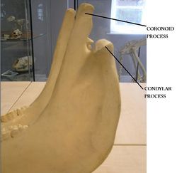
| − | + | [[Image:Jaw Articulation.jpg|thumb|right|250px|Jaw Articulation (horse) - Copyright RVC]] | |
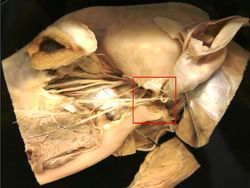
| + | [[Image:Temperomandibular Joint.jpg|thumb|right|250px|Temperomandibular Joint (dog) - Copyright RVC]] | ||
Mastication is the process whereby food is broken down by mechanical digestion in the [[Oral Cavity Overview - Anatomy & Physiology|oral cavity]]. The [[Cheeks|cheeks]] and [[Tongue - Anatomy & Physiology|tongue]] function to position food over the [[:Category:Teeth - Anatomy & Physiology|teeth]], where grinding can occur. Mastication requires correct muscle movements and jaw articulation. | Mastication is the process whereby food is broken down by mechanical digestion in the [[Oral Cavity Overview - Anatomy & Physiology|oral cavity]]. The [[Cheeks|cheeks]] and [[Tongue - Anatomy & Physiology|tongue]] function to position food over the [[:Category:Teeth - Anatomy & Physiology|teeth]], where grinding can occur. Mastication requires correct muscle movements and jaw articulation. | ||
| − | '''[[Rumination | + | '''[[Rumination|Rumination]]''' allows food to undergo mastication more than once. This is also called 'chewing the cud', it allows greater nutrients to be extracted and absorbed from the food particles. |
==Muscles of Mastication== | ==Muscles of Mastication== | ||
| − | |||
The muscles of mastication are well developed. | The muscles of mastication are well developed. | ||
| Line 18: | Line 19: | ||
All jaw closing muscles are derived from the first visceral arch and are innervated by the '''mandibular''' branch of the '''trigeminal''' nerve ([[Cranial Nerves - Anatomy & Physiology|CN V3]]). | All jaw closing muscles are derived from the first visceral arch and are innervated by the '''mandibular''' branch of the '''trigeminal''' nerve ([[Cranial Nerves - Anatomy & Physiology|CN V3]]). | ||
| − | The '''masseter muscle''' originates from the [[Skull and Facial Muscles - Anatomy & Physiology#Maxilla| | + | The '''masseter muscle''' originates from the [[Skull and Facial Muscles - Anatomy & Physiology#Maxilla|maxillary]] region of the skull and the zygomatic arch. It inserts on the wide area on the caudal side of the [[Skull and Facial Muscles - Anatomy & Physiology#Mandible (mandibula)|mandible]]. It has several divisions and causes '''unilateral''' and '''bilateral''' contraction. It also protrudes the jaw. |
The '''lateral pterygoid muscle''' originates from the [[Skull and Facial Muscles - Anatomy & Physiology#Pterygoid Bone (os pterygoideum)|pterygopalatine]] region of the skull. It inserts on the lateral aspect of the [[Skull and Facial Muscles - Anatomy & Physiology#Mandible (mandibula)|mandible]]. It also protrudes the jaw (one-sided contraction). | The '''lateral pterygoid muscle''' originates from the [[Skull and Facial Muscles - Anatomy & Physiology#Pterygoid Bone (os pterygoideum)|pterygopalatine]] region of the skull. It inserts on the lateral aspect of the [[Skull and Facial Muscles - Anatomy & Physiology#Mandible (mandibula)|mandible]]. It also protrudes the jaw (one-sided contraction). | ||
| Line 24: | Line 25: | ||
The '''medial pterygoid muscle''' originates from the [[Skull and Facial Muscles - Anatomy & Physiology#Pterygoid Bone (os pterygoideum)|pterygopalatine]] region of the skull. It inserts on the medial aspect of the [[Skull and Facial Muscles - Anatomy & Physiology#Mandible (mandibula)|mandible]]. It causes one-sided contraction to close the jaw. | The '''medial pterygoid muscle''' originates from the [[Skull and Facial Muscles - Anatomy & Physiology#Pterygoid Bone (os pterygoideum)|pterygopalatine]] region of the skull. It inserts on the medial aspect of the [[Skull and Facial Muscles - Anatomy & Physiology#Mandible (mandibula)|mandible]]. It causes one-sided contraction to close the jaw. | ||
| − | The '''temporal muscle''' originates from the lateral surface of the cranium. It inserts on the coronoid process. It pulls the [[Skull and Facial Muscles - Anatomy & Physiology#Mandible (mandibula)|mandible]] dorsally and also pulls the [[Skull and Facial Muscles - Anatomy & Physiology#Mandible (mandibula)|mandible]] rostrally (overbite) and caudally (underbite). | + | The '''temporal muscle''' originates from the lateral surface of the cranium. It inserts on the coronoid process. It pulls the [[Skull and Facial Muscles - Anatomy & Physiology#Mandible (mandibula)|mandible]] dorsally and also pulls the [[Skull and Facial Muscles - Anatomy & Physiology#Mandible (mandibula)|mandible]] rostrally (overbite) and caudally (underbite). |
===Lateral Translation of the Mandible=== | ===Lateral Translation of the Mandible=== | ||
| Line 31: | Line 32: | ||
==Jaw Articulation== | ==Jaw Articulation== | ||
| − | |||
| − | === | + | ===Temporomandibular Joint=== |
The articulation between the condylar process of the [[Skull and Facial Muscles - Anatomy & Physiology#Mandible (mandibula)|mandible]] and the mandibular process of the skull. It is a compartmentalised joint for rotational movement and lateral slide (grinding). It is a '''synovial joint'''. Caudal dislocation is prevented by a prominent retro-articular process (enlargement of the fossa). | The articulation between the condylar process of the [[Skull and Facial Muscles - Anatomy & Physiology#Mandible (mandibula)|mandible]] and the mandibular process of the skull. It is a compartmentalised joint for rotational movement and lateral slide (grinding). It is a '''synovial joint'''. Caudal dislocation is prevented by a prominent retro-articular process (enlargement of the fossa). | ||
| − | ===Mandibular | + | ===Mandibular Symphysis=== |
| − | |||
| − | Located at the rostral end of the [[Skull and Facial Muscles - Anatomy & Physiology#Mandible (mandibula)|mandible]]. It is a | + | Located at the rostral end of the [[Skull and Facial Muscles - Anatomy & Physiology#Mandible (mandibula)|mandible]]. It is a secondary cartilaginous joint between the left and right halves of the [[Skull and Facial Muscles - Anatomy & Physiology#Mandible (mandibula)|mandible]]. It is only found in dogs and ruminants. It has a precise occlusion and the [[Skull and Facial Muscles - Anatomy & Physiology#Mandible (mandibula)|Mandibular]] bones can move apart independently by rotation. It stops jaw breakages (Canid). |
==Species Differences== | ==Species Differences== | ||
===Hebivores=== | ===Hebivores=== | ||
| − | Herbivores have large '''masseter''' and '''pterygoid''' muscles for extensive chewing. | + | Herbivores have large '''masseter''' and '''pterygoid''' muscles for extensive chewing. Herbivorous species have a limited '''digastricus''' muscle. In the horse, the muscle insertion site for the '''masseter''' is large to snap jaw shut. |
===Carnivores=== | ===Carnivores=== | ||
| − | Carnivores have a large '''temporalis''' muscle for snapping the jaw shut, e.g. in lions and pitbull terriers. Canids have a larger ''' | + | Carnivores have a large '''temporalis''' muscle for snapping the jaw shut, e.g. in lions and pitbull terriers. Canids have a larger '''digastricus''' muscle than herbivores (but smaller in comparison with jaw closing muscles). In the dog, large forces are needed to shut the jaws, so the point of articulation of the '''temporomandibular joint''' is level with the teeth. |
==Links== | ==Links== | ||
| − | |||
| − | |||
| − | |||
'''Click here for [[Skull and Facial Muscles - Anatomy & Physiology]]''' | '''Click here for [[Skull and Facial Muscles - Anatomy & Physiology]]''' | ||
| − | + | {{Template:Learning | |
| − | + | |flashcards = [[Mastication Flashcards]]<br>[[Facial_Muscles_-_Musculoskeletal_-_Flashcards|Facial Muscles flashcards]] | |
| − | + | |videos = [http://stream2.rvc.ac.uk/Anatomy/canine/head_neck/Pot0220.mp4 Lateral surface of the head of a dog]<br>[http://stream2.rvc.ac.uk/Anatomy/canine/head_neck/Pot0258.mp4 Lateral section through the head of a dog] | |
| − | + | |dragster= [[Canine Head Skeletal Anatomy Resource (VI)]] | |
| − | + | }} | |
| − | |||
| + | ==Webinars== | ||
| + | <rss max="10" highlight="none">https://www.thewebinarvet.com/gastroenterology-and-nutrition/webinars/feed</rss> | ||
[[Category:Teeth - Anatomy & Physiology]] | [[Category:Teeth - Anatomy & Physiology]] | ||
[[Category:Musculoskeletal System - Anatomy & Physiology]] | [[Category:Musculoskeletal System - Anatomy & Physiology]] | ||
[[Category:Feeding Control]] | [[Category:Feeding Control]] | ||
[[Category:A&P Done]] | [[Category:A&P Done]] | ||
Latest revision as of 17:34, 7 November 2022
Overview
Mastication is the process whereby food is broken down by mechanical digestion in the oral cavity. The cheeks and tongue function to position food over the teeth, where grinding can occur. Mastication requires correct muscle movements and jaw articulation.
Rumination allows food to undergo mastication more than once. This is also called 'chewing the cud', it allows greater nutrients to be extracted and absorbed from the food particles.
Muscles of Mastication
The muscles of mastication are well developed.
Jaw Opening Muscles
The Digastricus muscle is the 'jaw opening' muscle. Its origin is the paracondylar process of the occipital bone. It inserts at the angle of the mandible. The muscle has two bellies; The caudal half from the second visceral arch innervated by the facial nerve (CN VII) and the cranial half from the first visceral arch, innervated by the mandibular branch of the trigeminal nerve (CN V3).
Jaw Closing Muscles
All jaw closing muscles are derived from the first visceral arch and are innervated by the mandibular branch of the trigeminal nerve (CN V3).
The masseter muscle originates from the maxillary region of the skull and the zygomatic arch. It inserts on the wide area on the caudal side of the mandible. It has several divisions and causes unilateral and bilateral contraction. It also protrudes the jaw.
The lateral pterygoid muscle originates from the pterygopalatine region of the skull. It inserts on the lateral aspect of the mandible. It also protrudes the jaw (one-sided contraction).
The medial pterygoid muscle originates from the pterygopalatine region of the skull. It inserts on the medial aspect of the mandible. It causes one-sided contraction to close the jaw.
The temporal muscle originates from the lateral surface of the cranium. It inserts on the coronoid process. It pulls the mandible dorsally and also pulls the mandible rostrally (overbite) and caudally (underbite).
Lateral Translation of the Mandible
The masseter muscle and the contralateral medial and lateral pterygoids are involved in the lateral translation of the mandible.
Jaw Articulation
Temporomandibular Joint
The articulation between the condylar process of the mandible and the mandibular process of the skull. It is a compartmentalised joint for rotational movement and lateral slide (grinding). It is a synovial joint. Caudal dislocation is prevented by a prominent retro-articular process (enlargement of the fossa).
Mandibular Symphysis
Located at the rostral end of the mandible. It is a secondary cartilaginous joint between the left and right halves of the mandible. It is only found in dogs and ruminants. It has a precise occlusion and the Mandibular bones can move apart independently by rotation. It stops jaw breakages (Canid).
Species Differences
Hebivores
Herbivores have large masseter and pterygoid muscles for extensive chewing. Herbivorous species have a limited digastricus muscle. In the horse, the muscle insertion site for the masseter is large to snap jaw shut.
Carnivores
Carnivores have a large temporalis muscle for snapping the jaw shut, e.g. in lions and pitbull terriers. Canids have a larger digastricus muscle than herbivores (but smaller in comparison with jaw closing muscles). In the dog, large forces are needed to shut the jaws, so the point of articulation of the temporomandibular joint is level with the teeth.
Links
Click here for Skull and Facial Muscles - Anatomy & Physiology
| Mastication Learning Resources | |
|---|---|
 Test your knowledge using drag and drop boxes |
Canine Head Skeletal Anatomy Resource (VI) |
 Test your knowledge using flashcard type questions |
Mastication Flashcards Facial Muscles flashcards |
 Selection of relevant videos |
Lateral surface of the head of a dog Lateral section through the head of a dog |
Webinars
Failed to load RSS feed from https://www.thewebinarvet.com/gastroenterology-and-nutrition/webinars/feed: Error parsing XML for RSS

