Difference between revisions of "Reabsorption and Secretion Along the Distal Tubule and Collecting Duct - Anatomy & Physiology"
| (46 intermediate revisions by 4 users not shown) | |||
| Line 1: | Line 1: | ||
| − | {{ | + | {{toplink |
| − | + | |backcolour = C1F0F6 | |
| + | |linkpage =Reabsorption and Secretion Along the Nephron - Anatomy & Physiology | ||
| + | |linktext =REABSORPTION AND SECRETION ALONG THE NEPHRON | ||
| + | |maplink = Urinary System (Content Map) - Anatomy & Physiology | ||
| + | |pagetype =Anatomy | ||
| + | }} | ||
| + | <br> | ||
| + | |||
==Distal Tubule== | ==Distal Tubule== | ||
| − | [[Image: | + | [[Image:disttubexch.jpg|right|thumb|300px|<small><center>Exchange in the Principal Cells of the Distal Tubule</center></small>]] |
* Important site of regulation of ions and water | * Important site of regulation of ions and water | ||
* Less emphasis on bulk transport compared with proximal tubule | * Less emphasis on bulk transport compared with proximal tubule | ||
| Line 8: | Line 15: | ||
* It is able to do this as it has high resistance epithelia. Allowing it to maintain substantial gradients across it | * It is able to do this as it has high resistance epithelia. Allowing it to maintain substantial gradients across it | ||
* Very important for the homeostasis of: | * Very important for the homeostasis of: | ||
| − | ** Sodium | + | ** [[Sodium Homeostasis - Physiology#Distal Tubule and Collecting Ducts| Sodium]] |
| − | ** Potassium | + | ** [[Potassium Homeostasis - Physiology#Distal Tubule| Potassium]] |
| − | ** Acid | + | ** [[Acid Base Balance By The Kidney - Anatomy & Physiology#Secretion of H+ in the Distal Tubule and Collecting Ducts| Acid Base]] |
| + | |||
| + | |||
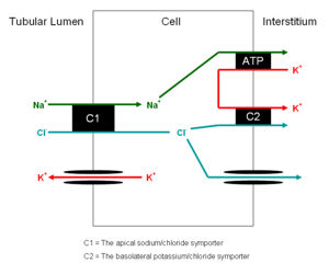
* There are two cell types present each with different functions. They are similar to the cells of the collecting ducts | * There are two cell types present each with different functions. They are similar to the cells of the collecting ducts | ||
** Principal cells | ** Principal cells | ||
*** Absorb sodium | *** Absorb sodium | ||
*** Excrete potassium and hydrogen | *** Excrete potassium and hydrogen | ||
| − | *** Site of action of [[ | + | *** Site of action of [[Aldosterone]] |
** Intercalated cells | ** Intercalated cells | ||
*** ATP driven proton secretion | *** ATP driven proton secretion | ||
===Juxtaglomerular Apparatus=== | ===Juxtaglomerular Apparatus=== | ||
| + | [[Image:juxtaapp.jpg|right|thumb|200px|<small><center>Histology section showing the juxtaglomerular apparatus (© RVC 2008)</center></small>]] | ||
* The terminal portion of the straight distal tubule contacts the afferent and efferent vessels supplying its own glomerulus | * The terminal portion of the straight distal tubule contacts the afferent and efferent vessels supplying its own glomerulus | ||
* These vessels are said to embrace the distal tubule | * These vessels are said to embrace the distal tubule | ||
* Here a special apparatus called the Juxtaglomerular Apparatus has 3 different structures: | * Here a special apparatus called the Juxtaglomerular Apparatus has 3 different structures: | ||
| − | ** The tubular epithelial cells of the distal tubule which are in contact with the arterioles supplying the glomerulus of that nephron are called the '''macula densa'''. They play a vital role in the [[ | + | ** The tubular epithelial cells of the distal tubule which are in contact with the arterioles supplying the glomerulus of that nephron are called the '''macula densa'''. They play a vital role in the [[Autoregulation of GFR - Anatomy and Physiology#Tubuloglomerular Feedback (TGF)|regulation of the GFR]]. |
| − | ** The Juxtaglomerular Cells are smooth muscle cells which adjoin the macula densa in the capillary wall. | + | ** The [[Juxtaglomerular Cells of The Distal Tubule - Renal Physiology | Juxtaglomerular Cells]] are smooth muscle cells which adjoin the macula densa in the capillary wall. |
** The Extraglomerular Mesangium has an unclear function | ** The Extraglomerular Mesangium has an unclear function | ||
| − | ==== | + | ===Developmental=== |
| − | |||
| − | |||
| − | |||
| − | |||
| − | |||
| − | |||
| − | |||
| − | |||
| − | |||
| − | |||
Develops from metanephric tubule | Develops from metanephric tubule | ||
==Collecting Duct== | ==Collecting Duct== | ||
| + | [[Image:collductloh.jpg|right|thumb|250px|<small><center>Histology section of the collecting duct showing the close proximity of the loop of henle (© RVC 2008)</center></small>]] | ||
[[Image:collductexch.jpg|right|thumb|250px|<small><center>Exchange in the Principal Cells of the Collecting Duct</center></small>]] | [[Image:collductexch.jpg|right|thumb|250px|<small><center>Exchange in the Principal Cells of the Collecting Duct</center></small>]] | ||
This part of the nephron has two cell types | This part of the nephron has two cell types | ||
| − | |||
===Principal Cells=== | ===Principal Cells=== | ||
| − | * [[Pituitary Gland - Anatomy & Physiology #Posterior Pituitary Gland | ADH]] acts on these cells inserting [[Aquaporins of the Kidney and Water Homeostasis - Anatomy & Physiology#What are Aquaporins|aquaporins]] into the cell membranes | + | |
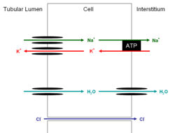
| − | * It is released from the [[Pituitary Gland - Anatomy & Physiology #Posterior Pituitary Gland | posterior pituitary gland]] | + | * [[Endocrine System - Pituitary Gland - Anatomy & Physiology #Posterior Pituitary Gland | ADH]] acts on these cells inserting [[Aquaporins of the Kidney and Water Homeostasis - Anatomy & Physiology#What are Aquaporins|aquaporins]] into the cell membranes |
| + | * It is released from the [[Endocrine System - Pituitary Gland - Anatomy & Physiology #Posterior Pituitary Gland | posterior pituitary gland]] | ||
===Intercalated cells=== | ===Intercalated cells=== | ||
| + | |||
* The intercalated cells can be subdivided further to: | * The intercalated cells can be subdivided further to: | ||
** Alpha intercalated cells secrete H<sup>+</sup> | ** Alpha intercalated cells secrete H<sup>+</sup> | ||
| Line 56: | Line 58: | ||
===Developmental=== | ===Developmental=== | ||
| + | |||
* Develops from branched ureteric bud | * Develops from branched ureteric bud | ||
===The Concentrating Mechanism, Aquaporins and ADH=== | ===The Concentrating Mechanism, Aquaporins and ADH=== | ||
| − | |||
| − | |||
| − | |||
| − | |||
| − | |||
| − | |||
| − | |||
| − | |||
| − | |||
| − | |||
| − | |||
| − | |||
| − | |||
| − | |||
| − | |||
| − | |||
| − | |||
| − | |||
| − | |||
| − | |||
| − | |||
| − | |||
| − | |||
| − | |||
| − | |||
| − | |||
| − | |||
| − | |||
| − | |||
| − | |||
| − | |||
| − | |||
| − | |||
| − | |||
| − | |||
| − | |||
| − | |||
| − | |||
| − | |||
| − | |||
| − | |||
| − | |||
| − | |||
| − | |||
| − | |||
| − | |||
| − | |||
| − | + | Water is drawn from the lumen of the tubule by the increasing hypertonicity of the surrounding tissue as the duct makes its way deeper into the medulla. However this reabsorption is only possible thanks to [[Aquaporins of the Kidney and Water Homeostasis - Anatomy & Physiology|ADH]] inserting [[Aquaporins of the Kidney and Water Homeostasis - Anatomy & Physiology#What are Aquaporins|aquaporins]] into the apical membrane. These channels are always present on the basolateral membrane of the epithelial cells but not on the apical membrane. The reabsorption would not be possible if the urine did not go back up the [[Loop Of Henle - Anatomy & Physiology #Thick ascending limb| thick ascending limb]] of the loop of henle, where its concentration was decreased by the reabsorption of salt, but instead went straight into the collecting ducts. Although this would mean very concentrated urine it would result in massive salt losses. Thus the collecting duct allows for very concentrated urine with minimal salt loss. It also allows the concentration of the urine to vary from dilute to concentrated under the control of the hypothalamus and ADH concentrations. | |
| − | [[ | ||
| − | [[ | ||
Revision as of 15:10, 3 September 2008
|
|
Distal Tubule
- Important site of regulation of ions and water
- Less emphasis on bulk transport compared with proximal tubule
- More emphasis on fine management
- It is able to do this as it has high resistance epithelia. Allowing it to maintain substantial gradients across it
- Very important for the homeostasis of:
- There are two cell types present each with different functions. They are similar to the cells of the collecting ducts
- Principal cells
- Absorb sodium
- Excrete potassium and hydrogen
- Site of action of Aldosterone
- Intercalated cells
- ATP driven proton secretion
- Principal cells
Juxtaglomerular Apparatus
- The terminal portion of the straight distal tubule contacts the afferent and efferent vessels supplying its own glomerulus
- These vessels are said to embrace the distal tubule
- Here a special apparatus called the Juxtaglomerular Apparatus has 3 different structures:
- The tubular epithelial cells of the distal tubule which are in contact with the arterioles supplying the glomerulus of that nephron are called the macula densa. They play a vital role in the regulation of the GFR.
- The Juxtaglomerular Cells are smooth muscle cells which adjoin the macula densa in the capillary wall.
- The Extraglomerular Mesangium has an unclear function
Developmental
Develops from metanephric tubule
Collecting Duct
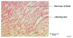
This part of the nephron has two cell types
Principal Cells
- ADH acts on these cells inserting aquaporins into the cell membranes
- It is released from the posterior pituitary gland
Intercalated cells
- The intercalated cells can be subdivided further to:
- Alpha intercalated cells secrete H+
- Beta intercalated cells secrete HCO3-
Developmental
- Develops from branched ureteric bud
The Concentrating Mechanism, Aquaporins and ADH
Water is drawn from the lumen of the tubule by the increasing hypertonicity of the surrounding tissue as the duct makes its way deeper into the medulla. However this reabsorption is only possible thanks to ADH inserting aquaporins into the apical membrane. These channels are always present on the basolateral membrane of the epithelial cells but not on the apical membrane. The reabsorption would not be possible if the urine did not go back up the thick ascending limb of the loop of henle, where its concentration was decreased by the reabsorption of salt, but instead went straight into the collecting ducts. Although this would mean very concentrated urine it would result in massive salt losses. Thus the collecting duct allows for very concentrated urine with minimal salt loss. It also allows the concentration of the urine to vary from dilute to concentrated under the control of the hypothalamus and ADH concentrations.


