Difference between revisions of "Canine Hindlimb - Anatomy & Physiology"
| (30 intermediate revisions by 8 users not shown) | |||
| Line 1: | Line 1: | ||
| − | {{ | + | {{toplink |
| + | |backcolour =CDE472 | ||
| + | |linkpage =Musculoskeletal System - Anatomy & Physiology | ||
| + | |linktext =Musculoskeletal System | ||
| + | |maplink = Musculoskeletal System (Content Map) - Anatomy & Physiology | ||
| + | |pagetype =Anatomy | ||
| + | |sublink1=Hindlimb - Anatomy & Physiology | ||
| + | |subtext1=HINDLIMB | ||
| + | }} | ||
| + | <br> | ||
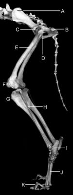
[[Image:Anatomy images bone promences dog Canine 6.jpeg|right|thumb|150px|'''The Canine Hindlimb Skeleton''']] | [[Image:Anatomy images bone promences dog Canine 6.jpeg|right|thumb|150px|'''The Canine Hindlimb Skeleton''']] | ||
| + | |||
| + | |||
==Pelvic Girdle and Hip== | ==Pelvic Girdle and Hip== | ||
| − | The pelvis encircles the pelvic cavity and has several functions | + | The pelvis encircles the pelvic cavity and has several functions; |
| − | + | * Protection of the pelvic viscera, most importantly the reproductive and urinary organs. | |
| − | + | * Essential in locomotion and posture | |
| + | * Forms the pelvic canal, the size of which can cause problems during parturition. | ||
| + | |||
===Bones=== | ===Bones=== | ||
| − | The pelvic girdle is formed by two hip bones which are joined ventrally at the cartilagenous '''pelvic symphysis''' and articulate dorsally with the sacrum. The three components of each hip bone are the [[ | + | |
| − | + | The pelvic girdle is formed by two hip bones which are joined ventrally at the cartilagenous '''pelvic symphysis''' and articulate dorsally with the sacrum. The three components of each hip bone are the [[Ilium - Anatomy and Physiology|ilium]], [[Pubis - Anatomy & Physiology|pubis]] and [[Ischium - Anatomy & Physiology|ischium]]. | |
| − | + | ||
| − | The bone that articulates with the hip bones to form the hip joint is the [[ | + | The bone that articulates with the hip bones to form the hip joint is the [[Femur - Anatomy & Physiology|Femur]]. |
| − | + | ||
| − | |||
====Canine Bone Specifics==== | ====Canine Bone Specifics==== | ||
| − | + | ||
| − | + | *'''[[Ilium - Anatomy and Physiology|Ilium]]''' | |
| − | + | **In the dog the '''tuber coxae''' isn't normally visible but is readily palpable. | |
| − | + | **The '''tuber coxae''' has two prominences; the cranial and caudal ventral iliac spines. | |
| − | + | **The '''sacral tuber''' has two prominences; the cranial and caudal dorsal iliac spines. | |
| − | + | **The '''iliac crest''' is wide and convex. | |
| − | + | **The '''ileal wing''' is orientated in an almost sagittal manner. | |
| − | + | ||
| − | + | *'''[[Ischium - Anatomy & Physiology|Ischium]]''' | |
| + | ** The '''ischial tuberosity''' is linear in shape. | ||
| + | |||
| + | *'''[[Femur - Anatomy & Physiology|Femur]]''' | ||
| + | ** The femoral head notch is circular and is situated in the centre of the head. | ||
| + | ** There is a distinct '''neck''' connecting the femoral head to the shaft. | ||
| + | ** The '''greater trochanter''' is level with the femoral head. | ||
===Joints and Synovial Structures=== | ===Joints and Synovial Structures=== | ||
| + | |||
====[[Hindlimb - Anatomy & Physiology#Sacroiliac Joint|Sacroiliac Joint]]==== | ====[[Hindlimb - Anatomy & Physiology#Sacroiliac Joint|Sacroiliac Joint]]==== | ||
| − | In dogs | + | |
| − | + | * In dogs the short branch of the '''dorsal sacroiliac ligaments''' connects the sacral tuberosity to the mamillary processes of the sacrum. | |
| + | * The '''sacrotuberous ligament''' consists of a fibrous cord between the iscial tuberosity and the transverse process of the last sacral vertebrae. (This ligament is absent in the cat.) | ||
| + | |||
| + | |||
====[[Hindlimb - Anatomy & Physiology#Coxafemoral|Coxafemoral/Hip Joint]]==== | ====[[Hindlimb - Anatomy & Physiology#Coxafemoral|Coxafemoral/Hip Joint]]==== | ||
| − | The dog has the greatest range of movement in this joint compared to other domestic species. It has the ability to flex, extend, rotate, adduct and abduct its whole limb because of this. | + | * The dog has the greatest range of movement in this joint compared to other domestic species. It has the ability to flex, extend, rotate, adduct and abduct its whole limb because of this. |
| − | |||
| − | |||
==Musculature== | ==Musculature== | ||
| − | |||
| − | |||
| − | |||
| − | |||
| − | |||
| − | |||
| − | |||
| − | The ''' | + | The muscles affecting the pelvic girdle and hip can be divided into two distinct groups: |
| + | |||
| + | *'''[[Girdle Musculature - Anatomy & Physiology|Girdle Musculature]]''' | ||
| + | ** '''Psoas Minor''' - a strong fleshy muscle. The tendon of insertion is bound to the iliac fascia and attaches to the arcuate line of the ilium. | ||
| + | ** '''Quadrate Lumbar''' - is stronger relative to other domestic species. It has both a thoracic and lumbar part. The thoracic part originates from the bodies of the last three thoracic vertebrae and inserts on the transverse processes of the cranial lumbar vertebrae. | ||
| + | |||
| + | *'''[[Rump Muscles - Anatomy & Physiology|Rump Muscles]]''' | ||
| + | ** '''Superficial Gluteal''' | ||
| + | *** Origin - gluteal fascia, lateral aspect of sacrum, sacral tuber of ilium, first caudal vertebra and the sacrotuberous ligament. | ||
| + | *** Insertion - via a tendon running caudodistally over the greater trochanter and inserting just distal to it. | ||
| + | ** '''Middle Gluteal''' | ||
| + | *** Origin - between the iliac crest and gluteal line of the ilium. | ||
| + | *** Insertion - Greater Trochanter | ||
| + | ** '''Tensor Muscle of the Fascia Lata''' | ||
| + | *** Origin - ventral aspect of iliac spine and aponeurosis of the middle gluteal. | ||
| + | *** Insertion - via the fascia lata to the patella. | ||
| + | *** Location - it fans out into the fascia lata and is bordered by the middlr gluteal dorsally and the sartorius muscle cranially. | ||
| + | ** '''Biceps''' | ||
| + | *** Origin - Cranial superficial head - sacrotuberous ligament. Caudal head - lateral aspect of iscial tuberosity | ||
| + | *** Insertion - The two muscle bellies unite at an aponeurosis attached to the stifle and crural fascia. This fascia then inserts on the patella, patella ligament, and tibial tuberosity. A distal tendon of the muscle seperates from the main muscle belly and passes under the adductor and along the gastrocnemius. It moves in front of the calcaneal tendon and combining with a tendon of the semitendinous muscle inserts on the calcaneal tuberosity. | ||
| + | ** '''Semitendinous''' | ||
| + | *** Origin - Caudal and ventrolateral aspect of the ischial tuberosity between the heads of the biceps and semimembranous muscle. | ||
| + | *** Insertion - via a strong tendon to the cranial aspect of the tendon. An accessory tendon, as mentioned above, also attaches to the calcaneal tuberosity. | ||
| + | ** '''Semimembranous''' | ||
| + | *** Origin - the ventral aspect of the ischium | ||
| + | *** Insertion - via a short tendon to the aponeurosis of the gastrocnemius and via a longer tendon to the medial femoral condyle and medial tibial condyle. | ||
| + | ** '''Sartorius''' | ||
| + | *** The cranial part originates on the iliac crest and unites with the femoral fascia and stifle fascia. | ||
| + | *** The caudal part originates from the ventral iliac spine and joins the aponeurosis of the gracilis on the cranial aspect of the tibia. | ||
| + | ** '''Pectineal''' | ||
| + | *** Origin - a fleshy origin from the iliopubic eminence and a tendinous origin from the prepubic tendon. | ||
| + | *** Insertion - via a tendinous attachment to the popliteal surface of the femur. | ||
| + | ** '''Adductor Muscles''' | ||
| + | *** The '''greater adductor muscle''' originates from the pelvic symphysis and prepubic tendon and inserts on the popliteal fossa and the lateral supracondylar tuberosity. | ||
| + | *** The '''short adductor''' originates on the pubic tubercle and inserts on the caudal aspect of the femur. | ||
| + | *** The '''long adductor''' is fused to the pectineal. (This is remains unfused in cats) | ||
| + | ** '''Internal Obturator''' | ||
| + | *** Origin - ischium, pubis and ischiatic arch. It covers the obturator foramen. | ||
| + | *** Insertion - Trochantic fossa via a strong tendon that passes over the lesser sciatic notch. | ||
==Proximal Hindlimb including Stifle and Tarsus== | ==Proximal Hindlimb including Stifle and Tarsus== | ||
===Bones=== | ===Bones=== | ||
| − | The bones immediately distal to the [[ | + | The bones immediately distal to the [[Femur - Anatomy & Physiology|Femur]] are the [[Hindlimb - Anatomy & Physiology#Tibia|Tibia]], [[Fibula - Anatomy & Physiology|Fibula]], [[Patella - Anatomy & Physiology|patella]] and some minor sesamoid bones. Some of these are involved in the stifle joint, weight-bearing and providing attachment for muscles. |
| − | Distal to these bones are the complex series of bones that make up the tarsus, these are the [[ | + | Distal to these bones are the complex series of bones that make up the tarsus, these are the [[Tarsal bones - Anatomy & Physiology|tarsal bones]] and [[Metatarsal bones - Anatomy & Physiology|metatarsals]]. |
====Canine Bone Specifics==== | ====Canine Bone Specifics==== | ||
| − | + | ||
| − | + | *'''[[Hindlimb - Anatomy & Physiology#Tibia|Tibia]]''' | |
| − | + | ** The cochlea has a lateral notch for the articulation with the [[fibula]]. | |
| − | + | ||
| − | + | *'''[[Fibula - Anatomy & Physiology|Fibula]]''' | |
| − | + | ** In the dog the fibula has maintained its entire length but still has reduced strength and function. | |
| − | + | ** The '''interosseous space''' seperates the [[Hindlimb - Anatomy & Physiology#Tibia|tibia]] from the fibula proximally and this gap is bridged by soft tissue. | |
| − | + | ** In thin dogs the entire bone is palpable but in heavy-set dogs only the proximal extremity is plalpable. | |
| − | + | ** The fibular head articulates with the lateral condyle of the [[Hindlimb - Anatomy & Physiology#Tibia|tibia]]. | |
| − | + | ||
| − | + | ||
| − | + | *'''[[Tarsal bones - Anatomy & Physiology#Talus|Talus]]''' | |
| + | ** The trochlea ridges are less pronounced and extend further distally than other species allowing for increased mobility. | ||
| + | ** The trochlea also articulates with the distal fibula and medial malleolus. | ||
| + | ** The body and head of the talus are seperated by a well defined '''neck'''. | ||
| + | |||
| + | |||
| + | *'''[[Tarsal bones - Anatomy & Physiology#Distal Row of Tarsal Bones| Distal Row of Tarsal Bones]]''' | ||
| + | ** The dog maintains the original number of 5 bones and has the central bone, 1st, 2nd, 3rd and 4th tarsal bones. | ||
=='''Joints and Synovial Structures'''== | =='''Joints and Synovial Structures'''== | ||
| − | '''[[ | + | |
| + | '''[[Stifle Joint - Anatomy & Physiology|Stifle Joint]]''' | ||
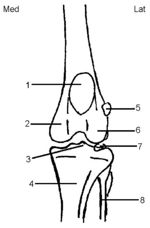
[[Image:stifle anatomy.jpg|thumb|right|150px|The Stifle, Cranial Caudal View - Copyright RVC]] | [[Image:stifle anatomy.jpg|thumb|right|150px|The Stifle, Cranial Caudal View - Copyright RVC]] | ||
| − | + | ||
| − | + | ||
| − | + | * Posesses a '''transverse ligament''' of the menisci. | |
| − | + | * The dog possesses one '''patellar ligament''' that is formed from the distal insertion of the quadriceps and connects the patella to the tibial tuberosity. It seperated from the joint capsule by the '''infrapatellar fat pad'''. There is often a synovial bursa between the distal part of the ligament and the tibial tuberosity. | |
| − | + | * The medial and lateral '''femeropatellar ligaments''' extend from the patellas to the femoral epicondyles and also have attachments to the '''fabella'''. These are two small sesamoid bones that are embedded in the head of the gastrocnemius muscle. | |
| − | + | * The joint capsule communicates directly with the femorotibial joint forming three sacs. One for the femeropatellar and one each for the medial and lateral femerotibial. These also contain the fabellae. The lateral pouch is extended to form the proximal tibiofibular joint capsule. | |
| + | |||
| + | |||
| + | '''[[Tarsal Joint/Hock - Anatomy & Physiology|Tarsal Joint/Hock]]''' | ||
| + | |||
| + | * Dogs have lateral movement as well as flexion and extension in their proximal intertarsal joints. | ||
| + | * The '''short medial collateral ligament''' has an extra branch in dogs that extends to the medial metatarsal bones. | ||
| + | |||
===Musculature=== | ===Musculature=== | ||
| − | + | ||
| − | + | '''[[Muscles of the Stifle - Anatomy & Physiology|Muscles of the Stifle]]''' | |
| − | + | * The division of the four parts of the quadriceps is less well defined than in other species. | |
| − | '''[[ | + | * The popliteal tendon of origin contains a sesamoid bone in carnivores. |
| + | |||
| + | '''[[Muscles of the Canine Crus - Anatomy & Physiology|Muscles of the Canine Crus]]''' | ||
| + | |||
| + | |||
==Vasculature of the Hindlimb== | ==Vasculature of the Hindlimb== | ||
| Line 87: | Line 157: | ||
'''[[Lymphatics of the Hindlimb - Anatomy & Physiology|Lymphatics of the Hindlimb]]''' | '''[[Lymphatics of the Hindlimb - Anatomy & Physiology|Lymphatics of the Hindlimb]]''' | ||
| − | |||
| − | |||
| − | |||
| − | |||
| − | |||
| − | |||
| − | |||
| − | |||
| − | |||
| − | |||
| − | |||
| − | |||
Revision as of 15:41, 15 September 2008
|
|
Pelvic Girdle and Hip
The pelvis encircles the pelvic cavity and has several functions;
- Protection of the pelvic viscera, most importantly the reproductive and urinary organs.
- Essential in locomotion and posture
- Forms the pelvic canal, the size of which can cause problems during parturition.
Bones
The pelvic girdle is formed by two hip bones which are joined ventrally at the cartilagenous pelvic symphysis and articulate dorsally with the sacrum. The three components of each hip bone are the ilium, pubis and ischium.
The bone that articulates with the hip bones to form the hip joint is the Femur.
Canine Bone Specifics
- Ilium
- In the dog the tuber coxae isn't normally visible but is readily palpable.
- The tuber coxae has two prominences; the cranial and caudal ventral iliac spines.
- The sacral tuber has two prominences; the cranial and caudal dorsal iliac spines.
- The iliac crest is wide and convex.
- The ileal wing is orientated in an almost sagittal manner.
- Ischium
- The ischial tuberosity is linear in shape.
- Femur
- The femoral head notch is circular and is situated in the centre of the head.
- There is a distinct neck connecting the femoral head to the shaft.
- The greater trochanter is level with the femoral head.
Joints and Synovial Structures
Sacroiliac Joint
- In dogs the short branch of the dorsal sacroiliac ligaments connects the sacral tuberosity to the mamillary processes of the sacrum.
- The sacrotuberous ligament consists of a fibrous cord between the iscial tuberosity and the transverse process of the last sacral vertebrae. (This ligament is absent in the cat.)
Coxafemoral/Hip Joint
- The dog has the greatest range of movement in this joint compared to other domestic species. It has the ability to flex, extend, rotate, adduct and abduct its whole limb because of this.
Musculature
The muscles affecting the pelvic girdle and hip can be divided into two distinct groups:
- Girdle Musculature
- Psoas Minor - a strong fleshy muscle. The tendon of insertion is bound to the iliac fascia and attaches to the arcuate line of the ilium.
- Quadrate Lumbar - is stronger relative to other domestic species. It has both a thoracic and lumbar part. The thoracic part originates from the bodies of the last three thoracic vertebrae and inserts on the transverse processes of the cranial lumbar vertebrae.
- Rump Muscles
- Superficial Gluteal
- Origin - gluteal fascia, lateral aspect of sacrum, sacral tuber of ilium, first caudal vertebra and the sacrotuberous ligament.
- Insertion - via a tendon running caudodistally over the greater trochanter and inserting just distal to it.
- Middle Gluteal
- Origin - between the iliac crest and gluteal line of the ilium.
- Insertion - Greater Trochanter
- Tensor Muscle of the Fascia Lata
- Origin - ventral aspect of iliac spine and aponeurosis of the middle gluteal.
- Insertion - via the fascia lata to the patella.
- Location - it fans out into the fascia lata and is bordered by the middlr gluteal dorsally and the sartorius muscle cranially.
- Biceps
- Origin - Cranial superficial head - sacrotuberous ligament. Caudal head - lateral aspect of iscial tuberosity
- Insertion - The two muscle bellies unite at an aponeurosis attached to the stifle and crural fascia. This fascia then inserts on the patella, patella ligament, and tibial tuberosity. A distal tendon of the muscle seperates from the main muscle belly and passes under the adductor and along the gastrocnemius. It moves in front of the calcaneal tendon and combining with a tendon of the semitendinous muscle inserts on the calcaneal tuberosity.
- Semitendinous
- Origin - Caudal and ventrolateral aspect of the ischial tuberosity between the heads of the biceps and semimembranous muscle.
- Insertion - via a strong tendon to the cranial aspect of the tendon. An accessory tendon, as mentioned above, also attaches to the calcaneal tuberosity.
- Semimembranous
- Origin - the ventral aspect of the ischium
- Insertion - via a short tendon to the aponeurosis of the gastrocnemius and via a longer tendon to the medial femoral condyle and medial tibial condyle.
- Sartorius
- The cranial part originates on the iliac crest and unites with the femoral fascia and stifle fascia.
- The caudal part originates from the ventral iliac spine and joins the aponeurosis of the gracilis on the cranial aspect of the tibia.
- Pectineal
- Origin - a fleshy origin from the iliopubic eminence and a tendinous origin from the prepubic tendon.
- Insertion - via a tendinous attachment to the popliteal surface of the femur.
- Adductor Muscles
- The greater adductor muscle originates from the pelvic symphysis and prepubic tendon and inserts on the popliteal fossa and the lateral supracondylar tuberosity.
- The short adductor originates on the pubic tubercle and inserts on the caudal aspect of the femur.
- The long adductor is fused to the pectineal. (This is remains unfused in cats)
- Internal Obturator
- Origin - ischium, pubis and ischiatic arch. It covers the obturator foramen.
- Insertion - Trochantic fossa via a strong tendon that passes over the lesser sciatic notch.
- Superficial Gluteal
Proximal Hindlimb including Stifle and Tarsus
Bones
The bones immediately distal to the Femur are the Tibia, Fibula, patella and some minor sesamoid bones. Some of these are involved in the stifle joint, weight-bearing and providing attachment for muscles.
Distal to these bones are the complex series of bones that make up the tarsus, these are the tarsal bones and metatarsals.
Canine Bone Specifics
- Fibula
- In the dog the fibula has maintained its entire length but still has reduced strength and function.
- The interosseous space seperates the tibia from the fibula proximally and this gap is bridged by soft tissue.
- In thin dogs the entire bone is palpable but in heavy-set dogs only the proximal extremity is plalpable.
- The fibular head articulates with the lateral condyle of the tibia.
- Talus
- The trochlea ridges are less pronounced and extend further distally than other species allowing for increased mobility.
- The trochlea also articulates with the distal fibula and medial malleolus.
- The body and head of the talus are seperated by a well defined neck.
- Distal Row of Tarsal Bones
- The dog maintains the original number of 5 bones and has the central bone, 1st, 2nd, 3rd and 4th tarsal bones.
Joints and Synovial Structures
- Posesses a transverse ligament of the menisci.
- The dog possesses one patellar ligament that is formed from the distal insertion of the quadriceps and connects the patella to the tibial tuberosity. It seperated from the joint capsule by the infrapatellar fat pad. There is often a synovial bursa between the distal part of the ligament and the tibial tuberosity.
- The medial and lateral femeropatellar ligaments extend from the patellas to the femoral epicondyles and also have attachments to the fabella. These are two small sesamoid bones that are embedded in the head of the gastrocnemius muscle.
- The joint capsule communicates directly with the femorotibial joint forming three sacs. One for the femeropatellar and one each for the medial and lateral femerotibial. These also contain the fabellae. The lateral pouch is extended to form the proximal tibiofibular joint capsule.
- Dogs have lateral movement as well as flexion and extension in their proximal intertarsal joints.
- The short medial collateral ligament has an extra branch in dogs that extends to the medial metatarsal bones.
Musculature
- The division of the four parts of the quadriceps is less well defined than in other species.
- The popliteal tendon of origin contains a sesamoid bone in carnivores.

