Difference between revisions of "Pedal Sepsis - Donkey"
Jump to navigation
Jump to search
m (Text replace - '{{review}}' to '') |
|||
| Line 1: | Line 1: | ||
| + | |||
| + | |||
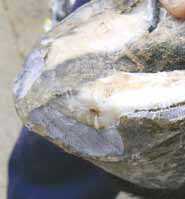
[[Image:Pedal sepsis donkey.jpg|right|thumb|200px|<small><center>Pedal sepsis (Image courtesy of [http://drupal.thedonkeysanctuary.org.uk The Donkey Sanctuary])</center></small>]] | [[Image:Pedal sepsis donkey.jpg|right|thumb|200px|<small><center>Pedal sepsis (Image courtesy of [http://drupal.thedonkeysanctuary.org.uk The Donkey Sanctuary])</center></small>]] | ||
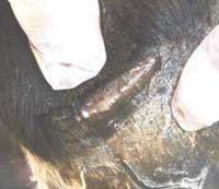
[[Image:Rupture at coronary band.jpg|right|thumb|200px|<small><center>Rupture at coronary band (Image courtesy of [http://drupal.thedonkeysanctuary.org.uk The Donkey Sanctuary])</center></small>]] | [[Image:Rupture at coronary band.jpg|right|thumb|200px|<small><center>Rupture at coronary band (Image courtesy of [http://drupal.thedonkeysanctuary.org.uk The Donkey Sanctuary])</center></small>]] | ||
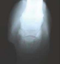
[[Image:Osteolytic lesion P3.jpg|right|thumb|200px|<small><center>Osteolytic lesion in the wing of distal phalanx (Image courtesy of [http://drupal.thedonkeysanctuary.org.uk The Donkey Sanctuary])</center></small>]] | [[Image:Osteolytic lesion P3.jpg|right|thumb|200px|<small><center>Osteolytic lesion in the wing of distal phalanx (Image courtesy of [http://drupal.thedonkeysanctuary.org.uk The Donkey Sanctuary])</center></small>]] | ||
| − | == | + | ==Description== |
The commonest cause of acute lameness, often presented as severe, non-weight-bearing. | The commonest cause of acute lameness, often presented as severe, non-weight-bearing. | ||
Revision as of 12:38, 18 March 2010


Description
The commonest cause of acute lameness, often presented as severe, non-weight-bearing.
Diagnosis
- Initially trim the hoof back to normal conformation - see routine trimming
- Expose a clear bearing surface free of all but deeper lesions
- Explore the entire weight-bearing surface, paying particular attention to the white line area, black marks, especially those adjacent to the sole axially, are suspicious.
- Hoof testers are of limited value, digital pressure at the coronary band may illicit a response from a related distal abscess.
Clinical Signs
- Abscesses frequently track proximally from the white line, eventually rupturing at the coronary band.
Treatment
Consider resecting the overlying hoof wall to facilitate drainage, but beware injury and prolapse of the underlying sensitive corium. An abaxial sesamoid nerve block will facilitate exploration.
The patient should be moved to food, water and shelter.
- Analgesia
- Tetanus prophylaxis
- Nursing care
- Sugardine mix is a cheap and effective final dressing application
- Potent antimicrobial action
- Promotes drying and hardening of the lesion
- Made by mixing Pevidine with granulated sugar to a crumbly mixture
- Beware prescribing systemic antibiotics prior to establishing appropriate drainage.
- 'Hot-tubbing' and flushing lesions with dilute povidone iodine (Pevidine antiseptic solution) are often useful.
- Protracted case may progress to involve the distal phalanx or result in extensive necrosis of the laminar corium. These cases require curettage under general anaesthesia. Glue-on plastic shoes are often beneficial and cost-effctive.
Prognosis
- Post-operative recovery after curettage may be protracted until the hoof capsule regenerates.
Sub-solar abscesses between the apex of the frog and the toe are often associated with terminal chronic foot disease or chronic laminitis and pedal bone degeneration.
References
- Crane, M. (2008) The donkey's foot In Svendsen, E.D., Duncan, J. and Hadrill, D. (2008) The Professional Handbook of the Donkey, 4th edition, Whittet Books, Chapter 10
|
|
This section was sponsored and content provided by THE DONKEY SANCTUARY |
|---|