Difference between revisions of "Category:Tongue - Pathology"
Jump to navigation
Jump to search
| Line 12: | Line 12: | ||
==Granulomatous and pyogranulomatous Inflammation== | ==Granulomatous and pyogranulomatous Inflammation== | ||
| − | === | + | ===[[Actinobacillosis]]=== |
| − | |||
| − | |||
| − | |||
| − | |||
| − | |||
| − | |||
| − | |||
| − | |||
| − | |||
| − | |||
| − | [[ | ||
| − | |||
| − | |||
| − | |||
| − | |||
| − | |||
| − | |||
| − | === | ||
| − | |||
| − | |||
| − | |||
| − | |||
| − | |||
| − | |||
| − | |||
| − | |||
| − | |||
| − | |||
| − | |||
==Eosinophilic Inflammation== | ==Eosinophilic Inflammation== | ||
Revision as of 13:09, 26 May 2010
Introduction
See anatomy and physiology of the tongue
Erosive & Ulcerative Pathology
Ulcerative glossitis
Vesicular Pathology
Neutrophilic Inflammation
Liquefactive necrosis
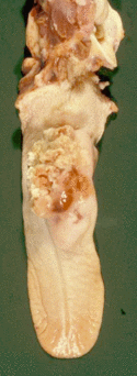
Granulomatous and pyogranulomatous Inflammation
Actinobacillosis
Eosinophilic Inflammation
Fibrinous/Diptheritic Inflammation
- Severe damage to epithelium produces exudation of fibrin -with formation of dry white fibrinous deposit - diptheritic membrane.
- Usually associated with organism fusobacterium necrophorum found everywhere in environment (but strict anaerobe).
- Produces lesions that damage epithelium due to toxin that damages vessels.
- Often a secondary invader but can be a primary pathogen.
Calf diptheria
- Caused by Fusiformis.
Clinical
- Seen in animals kept in cold, damp, muddy conditions.
- Usually associated with poor general health.
- Animals usually less than 6 months old, in groups and poorly kept.
- Lesions affect tongue, inside of mouth and larynx.
- Usually die fairly acutely as exudate blocks airway.
- May occur in young lambs secondary to orf (parapox) virus infection that can spread from lips to the inside of the mouth.
- (These lesions then become secondarily infected with Fusiformis.)
- Involvement of the pharynx and larynx may result in dyspnoea and the development of pneumonia.
- May lead to death.
Macroscopically
- Grossly, there is a firm swelling visible on the outer aspect of the cheek
- On the mucosal surface there is a deep, irregular ulcer, covered by a thick diphtheritic membrane.
Microscopically
- There is complete loss of the surface epithelium:
- a mass of necrotic debris
- polymorphs and fibrin is found overlying severely inflamed subepithelial tissues in which bacterial colonies may be seen and around which there may be marked fibrosis in an attempt to “wall off” the lesion.
Haemorrhagic Inflammation
- Complete loss of integrity of epithelium. Uncommon.
- Characteristic of “Blue Tongue”, a Reovirus infection of sheep - (v. rare – doesn’t occur in UK !).
- Epithelium lost and haemorrhage produces blue / black discoloration of the tongue, hence the name.
Tongue Lymphoma
See also Tongue Trauma Clinical Page
Fungal
- Fungal infections are relatively rare but do occur.
- The best known is infection with the yeast Candida albicans which causes thrush.
Pages in category "Tongue - Pathology"
The following 10 pages are in this category, out of 10 total.
