Difference between revisions of "Oesophagitis"
TestStudent (talk | contribs) |
JamesSwann (talk | contribs) |
||
| (20 intermediate revisions by 4 users not shown) | |||
| Line 1: | Line 1: | ||
| − | {{ | + | {{unfinished}} |
| − | == | + | |
| − | [[Image:Oesophagitis.jpg|thumb|right|250px| | + | |
| − | Oesophagitis refers to [[Inflammation - Pathology|inflammation]] of the oesophagus. This usually involves the '''mucosa''' but can involve the deeper layers of the submucosa and muscularis and it may follow an '''acute''' or '''chronic''' course. The oesophagus is usually protected from physical or chemical damage by mucus (produced by simple tubuloacinar glands along its whole length in dogs and in the rostral portion in cats) | + | ==Description== |
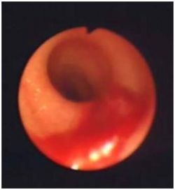
| + | [[Image:Oesophagitis.jpg|thumb|right|250px|Oesophagitis - Copyright David Walker RVC]] | ||
| + | Oesophagitis refers to [[Inflammation - Pathology|inflammation]] of the oesophagus. This usually involves the '''mucosa''' but can involve the deeper layers of the submucosa and muscularis and it may follow an '''acute''' or '''chronic''' course. The oesophagus is usually protected from physical or chemical damage by mucus (produced by simple tubuloacinar glands along its whole length in dogs and in the rostral portion in cats) and by peristaltic waves and the upper and lower oesophageal sphincters which prevent ingesta or regurgitated material from remaining in contact with the oesophageal wall. Oesophagitis is a serious condition and, if not treated, it may progress to ulceration, rupture, [[Oesophageal Stricture|stricture formation]] or derangement of normal motility ([[Megaoesophagus|megaoesophagus]]). The most common causes are: | ||
*'''Physical Injury''' | *'''Physical Injury''' | ||
**Ingestion of [[Oesophageal Foreign Body|'''foreign bodies''']] which lodge in the oesophagus. | **Ingestion of [[Oesophageal Foreign Body|'''foreign bodies''']] which lodge in the oesophagus. | ||
| Line 9: | Line 11: | ||
**'''Gastro-oesophageal reflux''', which may occur with '''general anaesthesia''' or [[Hiatal Hernia|'''hiatal hernias''']]. | **'''Gastro-oesophageal reflux''', which may occur with '''general anaesthesia''' or [[Hiatal Hernia|'''hiatal hernias''']]. | ||
**'''Chronic vomiting''' | **'''Chronic vomiting''' | ||
| − | **Ingestion of '''caustic''' or '''irritant substances''', including ''' | + | **Ingestion of '''caustic''' or '''irritant substances''', including '''doxycycline''' in cats. |
==Signalment== | ==Signalment== | ||
| − | Any age group can be affected and there is usually a history suggestive of a particular cause, such as a recent general anaesthetic or administration of | + | Any age group can be affected and there is usually a history suggestive of a particular cause, such as a recent general anaesthetic or administration of doxycylcine to a cat. |
==Diagnosis== | ==Diagnosis== | ||
| Line 25: | Line 27: | ||
The results of diagnostic tests are usually unremarkable but, as with any inflammatory process, there may be an overall '''leucocytosis''' caused by '''neutrophilia''' in the acute stage. | The results of diagnostic tests are usually unremarkable but, as with any inflammatory process, there may be an overall '''leucocytosis''' caused by '''neutrophilia''' in the acute stage. | ||
===Diagnostic Imaging=== | ===Diagnostic Imaging=== | ||
| − | ''' | + | '''Survey radiographs''': generally normal. Signs of aspiration pneumonia may be seen in ventral lung lobes. It can be better differentiated using barium-contrast studies which may show: |
| − | + | *an irregular mucosal surface | |
| − | + | *narrowing | |
| − | '''Endoscopy''' | + | *a dilated oesophagus |
| − | + | *oesophageal hypomotility | |
| + | '''Endoscopy''': To better diagnose oesophagitis, endoscopy with biopsies can be used. Severe oesophagitis will be seen with an oedematous mucosa that is hyperaemic, ulcerated and actively bleeding, whilst less severe cases may require several mucosal biopsies to diagnose the condition. | ||
==Treatment== | ==Treatment== | ||
| − | + | Mild oesophagitis: | |
| − | * | + | *withdraw oral food for 2-3 days and manage as an outpatient. |
| − | * | + | More severe oesophagitis: |
| − | * | + | *may need admitting to the hospital, Nil Per Os and animal may require enteral or parenteral nutritional support. |
| − | + | Drugs: | |
| − | * | + | *oral sucralfate suspension |
| − | * | + | *gastric acid secretory inhibitors (e.g. ranitidine, omeprazole) can be useful in cases of gastro-oesophageal reflux |
| − | + | *broad spectrum antibiotics in animals with sever oesophagitis or aspiration pneumonia | |
| + | *analgesics | ||
==Prognosis== | ==Prognosis== | ||
| − | Mild oesophagitis has a good prognosis | + | Mild oesophagitis has a good prognosis whereas ulcerative oesophagitis and animals suffering from aspiration pneumonia have a more guarded prognosis. |
| + | ==References== | ||
| − | |||
| − | |||
| − | |||
| − | |||
| − | |||
| − | |||
Hall, E.J, Simpson, J.W. and Williams, D.A. (2005) '''BSAVA Manual of Canine and Feline Gastroenterology (2nd Edition)''' ''BSAVA'' | Hall, E.J, Simpson, J.W. and Williams, D.A. (2005) '''BSAVA Manual of Canine and Feline Gastroenterology (2nd Edition)''' ''BSAVA'' | ||
| − | + | Merck & Co (2008) '''The Merck Veterinary Manual''' | |
| − | |||
| − | Merck & Co (2008) '''The Merck Veterinary Manual''' | ||
| − | |||
| − | |||
| − | |||
| − | |||
| − | |||
| − | |||
[[Category:Oesophagus_-_Pathology]] | [[Category:Oesophagus_-_Pathology]] | ||
| − | [[Category: | + | [[Category:To_Do_-_James]] |
| − | |||
Revision as of 10:14, 7 July 2010
| This article is still under construction. |
Description
Oesophagitis refers to inflammation of the oesophagus. This usually involves the mucosa but can involve the deeper layers of the submucosa and muscularis and it may follow an acute or chronic course. The oesophagus is usually protected from physical or chemical damage by mucus (produced by simple tubuloacinar glands along its whole length in dogs and in the rostral portion in cats) and by peristaltic waves and the upper and lower oesophageal sphincters which prevent ingesta or regurgitated material from remaining in contact with the oesophageal wall. Oesophagitis is a serious condition and, if not treated, it may progress to ulceration, rupture, stricture formation or derangement of normal motility (megaoesophagus). The most common causes are:
- Physical Injury
- Ingestion of foreign bodies which lodge in the oesophagus.
- Passage of nasogastric or pharyngostomy feeding tubes or of large endoscopes.
- Chemical Injury
- Gastro-oesophageal reflux, which may occur with general anaesthesia or hiatal hernias.
- Chronic vomiting
- Ingestion of caustic or irritant substances, including doxycycline in cats.
Signalment
Any age group can be affected and there is usually a history suggestive of a particular cause, such as a recent general anaesthetic or administration of doxycylcine to a cat.
Diagnosis
Clinical Signs
Signs include:
- Regurgitation and hypersalivation/ptyalism are the most common signs. Regurgitation can be recognised if the animal shows no abdominal effort (as would occur with vomiting) and if the material produced still resembles the food that was eaten with little apparent digestion. The material produced is often covered with white foam but bile should never be present.
- Animals may appear to have difficulty in swallowing and often appear to make multiple swallowing efforts and to extend their necks during swallowing.
- Animals may show signs of pain during swallowing (odynophagia) and may therefore become anorexic.
- Like any animals which regurgitate repeatedly, those with oesophagitis may develop aspiration pneumonia and show signs of pyrexia, coughing, tachypnoea and dyspnoea.
Laboratory Tests
The results of diagnostic tests are usually unremarkable but, as with any inflammatory process, there may be an overall leucocytosis caused by neutrophilia in the acute stage.
Diagnostic Imaging
Survey radiographs: generally normal. Signs of aspiration pneumonia may be seen in ventral lung lobes. It can be better differentiated using barium-contrast studies which may show:
- an irregular mucosal surface
- narrowing
- a dilated oesophagus
- oesophageal hypomotility
Endoscopy: To better diagnose oesophagitis, endoscopy with biopsies can be used. Severe oesophagitis will be seen with an oedematous mucosa that is hyperaemic, ulcerated and actively bleeding, whilst less severe cases may require several mucosal biopsies to diagnose the condition.
Treatment
Mild oesophagitis:
- withdraw oral food for 2-3 days and manage as an outpatient.
More severe oesophagitis:
- may need admitting to the hospital, Nil Per Os and animal may require enteral or parenteral nutritional support.
Drugs:
- oral sucralfate suspension
- gastric acid secretory inhibitors (e.g. ranitidine, omeprazole) can be useful in cases of gastro-oesophageal reflux
- broad spectrum antibiotics in animals with sever oesophagitis or aspiration pneumonia
- analgesics
Prognosis
Mild oesophagitis has a good prognosis whereas ulcerative oesophagitis and animals suffering from aspiration pneumonia have a more guarded prognosis.
References
Hall, E.J, Simpson, J.W. and Williams, D.A. (2005) BSAVA Manual of Canine and Feline Gastroenterology (2nd Edition) BSAVA
Merck & Co (2008) The Merck Veterinary Manual
