Difference between revisions of "Otodectes cynotis"

From WikiVet English
Jump to navigation Jump to search
(20 intermediate revisions by 4 users not shown)
Line 1: Line 1:
{{OpenPagesTop}}
+
{{unfinished}}
{{Taxobox
 
|name              =''Otodectes cynotis''
 
|phylum            =Arthropoda
 
|class              =[[:Category:Arachnida|Arachnida]]
 
|order              =Astigmata
 
|family            =Psoroptidia
 
|genus              =Otodectes
 
|species            =''O.cynotis''
 
}}
 
Also known as: '''''Ear mite'''
 
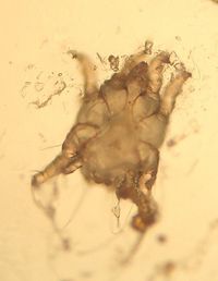
[[File:Otodectes cynotis.jpg|thumb|200px|right|''Otodectes cynotis'' C. Antonczyk 2008]]
 
[[File:Otodectes.jpg|thumb|200px|right|''Otodectes cynotis'' Caroldermoid 2006, wikimedia commons]]
 
  
==Introduction==
+
[[File:Otodectes cynotis.jpg|thumb|200px|right|Otodectes cynotis (Courtesy of C. Antonczyk)]]
''Otodectes cynotis'' mites are [[Non-Burrowing Mites|surface mites]]. They are the cause of [[Otitis Externa - Cat and Dog|otodectic otitis]], the most common mange of dogs and cats in the world. They are also found in the fox and the ferret. The mites inhabit the inner ear and feed on ear debris, they appear white in colour.
 
  
Pruritus is caused by irritation and the saliva of the mites, which is immunogenic. '''Secondary bacterial infection''' is common, resulting in purulent otitis externa which will require more treatment.
+
Otodectes cynotis mites are [[Non-Burrowing Mites|surface mites]]. They are the cause of [[Otodectic Dermatosis|otodectic skin infestation]], the most common mange of dogs and cats in the world. They are also found in the fox and the ferret. The mites inhabit the inner ear and feed on ear debris.
 
 
In cats, another presentation can be seen, which is that of an ectopic infection where signs are seen on other body parts such as the tail. This is due to contact e.g. cats sleeping in a curled position would allow mites to infest that area. Transmission of the mites can occur to kittens whilst they are suckling.


==Identification==
 
==Identification==
The mites have closed keratinous bars, '''apodemes''' on their ventral surface. They are smaller in size than [[psoroptes cuniculi]] and have short pedicles on their first and second pairs of legs.
 
 
==Lifecycle==
 
The Life cycle of an ''Otodectes'' mite takes '''3 weeks'''. The females lay around five eggs a day on the surface of the ear canal. Four days later, larvae hatch and become nymphs. There are two nymphal stages before an adult mite is formed.
 
 
==Clinical Signs==
 
Clinical signs include '''head shaking, scratching of the ear''' and even the development of an '''aural haematoma''' because of the two above signs. The animal may often resent this area being touched.
 
 
A '''brown waxy exudate''' is produced and this later becomes crusty. If secondary bacterial infection has occurred then pus will also be seen in the ear canal.
 
  
==Diagnosis==
+
The mites have closed keratinous bars ('''apodemes''') on their ventral surface. They are smaller in size than [[psoroptes cuniculi]]
Clinical signs and history are indicative of the disease.
 
  
'''Visualisation''' of the mites via an auroscope will provide definitive diagnosis.
+
*Life cycle takes '''3 weeks'''
  
== Treatment ==
 
Topical ear drops usually contain '''acaricide, fungicide, antibiotics and steroids'''. These should be dropped into the ear and the base of the ear then massaged to help the drops disperse. In cases of severe wax build up, ceruminolytics may be useful.
 
  
'''Selamectin''' as a spot on also acts to prevent ''Otodectes''.
 
  
'''All in contact animals should be treated''' in the household as these may be asymptomatic carriers.
+
'''Pathogenesis'''
 +
*The majority of cats harbour the mites, however only a few show symptoms
 +
**Transmission occurs whilst kittens are suckling
  
== References ==
+
*Common cause of [[Otitis Externa - Small Animal|otitis externa]] in dogs
Blood, D.C. and Studdert, V. P. (1999) '''Saunders Comprehensive Veterinary Dictionary''' (2nd Edition),'' Elsevier Science''.
 
<br>
 
Bond, R. (2008) '''Dermatology Study Guide,''''' Royal Veterinary College.''
 
<br>
 
Foster, A, and Foll, C. (2003) '''BSAVA small animal dermatology''' (second edition), ''British Small Animal Veterinary Association ''
 
<br>
 
Fox, M and Jacobs, D. (2007) '''Parasitology Study Guide Part 1: Ectoparasites, '''''Royal Veterinary College ''
 
  
 +
*Brown waxy exudate produced
  
<big><b>Also see:
+
*Can lead to secondary infection
:[[Otitis Externa - Cat and Dog|Ear Mites in Cats and Dogs]]
 
:[[Mites - Ferrets#Ear Mites|Ear Mites in Ferrets]]
 
</b></big>
 
  
{{Learning
+
*Clinical signs are apparent
|flashcards = [[Mites_Flashcards|Mites Flashcards]]
+
**Head shaking
|literature search = [http://www.cabdirect.org/search.html?rowId=1&options1=AND&q1=%22Otodectes+cynotis%22&occuring1=title&rowId=2&options2=AND&q2=&occuring2=freetext&rowId=3&options3=AND&q3=&occuring3=freetext&x=67&y=17&publishedstart=yyyy&publishedend=yyyy&calendarInput=yyyy-mm-dd&la=any&it=any&show=all ''Otodectes cynotis'' publications]
+
**Ear scratching
|Vetstream = [https://www.vetstream.com/canis/search?s=mite Mites]
+
**Aural haematomata
}}


 +
'''Treatment'''
 +
*Acaracidal ear drops
 +
**Massage base of ear to disperse drops after treatment
  
 +
*Most treatments need to be repeated in 10-14 days to kill newly hatched mites
  
{{review}}
+
*Selamectin can be used as a spot-on treatment
 +
**Prolonged duration of action
  
{{OpenPages}}
+
*Treat all in-contact animals
 +
**These may be asymptomatic carriers
 +
[[Category:Non-Burrowing_Mites]][[Category:Dog]]
  
[[Category:Non-Burrowing_Mites]]
+
[[Category:To_Do_-_AimeeHicks]]
[[Category:Expert_Review]]
 

Revision as of 07:00, 11 July 2010


Otodectes cynotis (Courtesy of C. Antonczyk)

Otodectes cynotis mites are surface mites. They are the cause of otodectic skin infestation, the most common mange of dogs and cats in the world. They are also found in the fox and the ferret. The mites inhabit the inner ear and feed on ear debris.

Identification

The mites have closed keratinous bars (apodemes) on their ventral surface. They are smaller in size than psoroptes cuniculi

  • Life cycle takes 3 weeks


Pathogenesis

  • The majority of cats harbour the mites, however only a few show symptoms
    • Transmission occurs whilst kittens are suckling
  • Brown waxy exudate produced
  • Can lead to secondary infection
  • Clinical signs are apparent
    • Head shaking
    • Ear scratching
    • Aural haematomata


Treatment

  • Acaracidal ear drops
    • Massage base of ear to disperse drops after treatment
  • Most treatments need to be repeated in 10-14 days to kill newly hatched mites
  • Selamectin can be used as a spot-on treatment
    • Prolonged duration of action
  • Treat all in-contact animals
    • These may be asymptomatic carriers