Difference between revisions of "Intestinal Arterial Thromboembolism"
Jump to navigation
Jump to search
| (11 intermediate revisions by the same user not shown) | |||
| Line 10: | Line 10: | ||
** Leads to vasoconstriction | ** Leads to vasoconstriction | ||
*** May occlude lumen and encourage thromboemboli. | *** May occlude lumen and encourage thromboemboli. | ||
| − | ** Can cause ischaemic necrosis of a segment of [[Small Intestine | + | ** Can cause ischaemic necrosis of a segment of [[Small Intestine - Anatomy & Physiology|small intestine]] |
** Is less common now that [[Strongylus vulgaris|'''''Strongylus vulgaris''''']] infections are declining. | ** Is less common now that [[Strongylus vulgaris|'''''Strongylus vulgaris''''']] infections are declining. | ||
* E.g. '''equine''' [[Salmonellosis|'''salmonellosis''']]. | * E.g. '''equine''' [[Salmonellosis|'''salmonellosis''']]. | ||
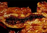
[[Image:Infaction of the small bowel.jpg|thumb|right|150px|Infarction of the small bowel (Courtesy of Bristol BioMed Image Archive)]] | [[Image:Infaction of the small bowel.jpg|thumb|right|150px|Infarction of the small bowel (Courtesy of Bristol BioMed Image Archive)]] | ||
| − | |||
| − | |||
| − | |||
| − | |||
| − | |||
| − | |||
| − | |||
| − | |||
| − | |||
| − | |||
===Small Animals=== | ===Small Animals=== | ||
| Line 37: | Line 27: | ||
* Similar to that caused by venous congestion. | * Similar to that caused by venous congestion. | ||
* See sharply delineated dark areas in bowel that are flaccid with loss of tone. | * See sharply delineated dark areas in bowel that are flaccid with loss of tone. | ||
| − | ** These become necrotic followed later by peritonitis. | + | ** These become necrotic followed later by peritonitis.[[Category:Intestine_-_Vascular_Disturbances]] |
| − | + | [[Category:Dog]][[Category:Cat]] | |
| − | |||
| − | [[Category:Intestine_-_Vascular_Disturbances]] | ||
| − | [[Category: | ||
[[Category:To_Do_-_Clinical]] | [[Category:To_Do_-_Clinical]] | ||
| − | [[Category: | + | [[Category:Cardiovascular_Disorders_-_Horse]] |
| − | [[Category: | + | [[Category:Small_Intestinal_Disorders_-_Horse]] |
| − | |||
Revision as of 13:30, 30 July 2010
- Non-strangulation infarction.
- There is often a functional obstruction at point of infarction.
- Relatively rare as the bowel has a good anastomosing blood supply.
Horses
- E.g. Strongylus vulgaris larvae migrating in cranial mesenteric artery in horse
- Cause arteritis with thickening of wall
- Due to fibrin and debris deposition and hypersensitivity reaction
- Leads to vasoconstriction
- May occlude lumen and encourage thromboemboli.
- Can cause ischaemic necrosis of a segment of small intestine
- Is less common now that Strongylus vulgaris infections are declining.
- Cause arteritis with thickening of wall
- E.g. equine salmonellosis.
Small Animals
- Especially dogs
- Road traffic accidents produce and infact in the gut.
- Renal disease also causes infarction.
- Particularly nephrotic syndrome.
- Anticoagulant proteins are lost in the urine, leading to a prothrombic state in the ciruclation.
Pathology
- Similar to that caused by venous congestion.
- See sharply delineated dark areas in bowel that are flaccid with loss of tone.
- These become necrotic followed later by peritonitis.

