Difference between revisions of "Myxomatosis"
| (13 intermediate revisions by 2 users not shown) | |||
| Line 1: | Line 1: | ||
| − | {{ | + | {{unfinished}} |
| − | |||
| − | |||
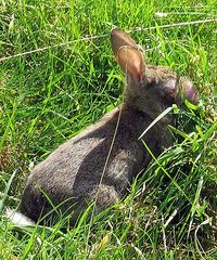
| − | == | + | [[Image:Myxomatosis_rabbit_logo.jpg|200px|right|thumb|A European Rabbit afflicted by Myxomatosis <br> Chris Bayley, WikiMedia Commons]] |
| + | |||
| + | ==Description== | ||
Myxomatosis is a highly contagious viral condition of rabbits caused by the myxoma virus, a member of the poxvirus group. It was first recognised in the UK in 1953 after it crossed the channel from France where it was illegally introduced in 1952. It is carried mainly by arthropods, particularly the rabbit flea, ''Spilopsyllus cuniculi''. The disease is also transmitted by direct or indirect contact with ocular or skin discharges or by mechanical vectors. The disease is characterised by subcutaneous mucinous lesions and nodular tumours and is associated with a high mortality rate. | Myxomatosis is a highly contagious viral condition of rabbits caused by the myxoma virus, a member of the poxvirus group. It was first recognised in the UK in 1953 after it crossed the channel from France where it was illegally introduced in 1952. It is carried mainly by arthropods, particularly the rabbit flea, ''Spilopsyllus cuniculi''. The disease is also transmitted by direct or indirect contact with ocular or skin discharges or by mechanical vectors. The disease is characterised by subcutaneous mucinous lesions and nodular tumours and is associated with a high mortality rate. | ||
| Line 11: | Line 11: | ||
The myxoma virus infects several cell types including mucosal cells, lymphocytes and fibroblasts. In addition to primary and secondary tumour development, there is severe immunosuppression leading to overwhelming infections by opportunistic gram-negative bacteria particularly affecting the conjunctiva and nasal passages. | The myxoma virus infects several cell types including mucosal cells, lymphocytes and fibroblasts. In addition to primary and secondary tumour development, there is severe immunosuppression leading to overwhelming infections by opportunistic gram-negative bacteria particularly affecting the conjunctiva and nasal passages. | ||
| − | Virus multiplication and tumour-like lesions occur initially at the site of intradermal inoculation. This is followed by spread to regional lymph nodes and cell-associated viraemia, with generalization to the skin and internal organs. Gelatinous proliferative nodules develop all over the body, especially at orifices such as the eyes, anus, nose. | + | Virus multiplication and tumour-like lesions occur initially at the site of intradermal inoculation. This is followed by spread to regional lymph nodes and cell-associated viraemia, with generalization to the skin and internal organs. Gelatinous proliferative nodules develop all over the body, especially at orifices such as the eyes, anus, nose. The rabbit usually dies within 12 days. |
| − | |||
| − | |||
| − | |||
| − | |||
| − | |||
| − | |||
==Clinical signs== | ==Clinical signs== | ||
| Line 30: | Line 24: | ||
==Prevention== | ==Prevention== | ||
| − | Vaccination and control of insect parasites are the most important means of disease prevention in domestic rabbits. In order to control fleas, wild rabbits should be kept away from pet rabbits and spot-on products may be used. Mosquito control can be achieved using insect repellent strips and fine mesh netting | + | Vaccination and control of insect parasites are the most important means of disease prevention in domestic rabbits. In order to control fleas, wild rabbits should be kept away from pet rabbits and spot-on products may be used. Mosquito control can be achieved using insect repellent strips and fine mesh netting. |
| − | The myxomatosis vaccine currently used in the UK is a live vaccine containing ''Shope fibroma'' virus (Nobivac Myxo, Intervet) | + | The myxomatosis vaccine currently used in the UK is a live vaccine containing ''Shope fibroma'' virus (Nobivac Myxo, Intervet). Antibodies made against ''Shope fibroma'' provide cross immunity against myxomatosis. Intradermal vaccination is performed in order to achieve adequate immunity and annual booster vaccination is recommended. Live attenuated vaccines have been used elsewhere in Europe but have been associated with other side effects such as immunosuppression. |
| − | |||
| − | |||
| − | |||
| − | |||
| − | |||
==Treatment== | ==Treatment== | ||
| − | Although the mortality rate in affected rabbits is high, recovery is possible and | + | Although the mortality rate in affected rabbits is high, recovery is possible. High ambient temperatures have been reported to increase recovery rates and a warm environment should be provided. Antibiotics and non-steroidal anti-inflammatories are commonly used to treat the disease, however corticosteroids are contraindicated due to their immunosuppressive effects. |
| − | |||
| − | |||
| − | |||
| − | |||
| − | |||
| − | |||
| − | |||
| − | |||
| − | |||
| − | |||
| − | |||
| − | |||
| − | |||
| − | |||
| − | |||
==References== | ==References== | ||
| Line 61: | Line 35: | ||
*Harcourt-Brown, F. (2002) '''Textbook of Rabbit Medicine''' ''Elsevier Health Sciences'' | *Harcourt-Brown, F. (2002) '''Textbook of Rabbit Medicine''' ''Elsevier Health Sciences'' | ||
*Kayne, S. B., Jepson, M. H. (2004) '''Veterinary Pharmacy''' ''Pharmaceutical Press'' | *Kayne, S. B., Jepson, M. H. (2004) '''Veterinary Pharmacy''' ''Pharmaceutical Press'' | ||
| − | |||
| − | |||
| − | |||
| − | |||
| − | |||
| − | |||
| − | |||
| + | [[Category:Leporipoxviruses]][[Category:Rabbit Viruses]] | ||
| − | + | [[Category:To_Do_-_SophieIgnarski]] | |
| − | + | [[Category:To_Do_-_Review]] | |
| − | |||
| − | [[Category: | ||
Revision as of 13:45, 29 September 2010
| This article is still under construction. |
Description
Myxomatosis is a highly contagious viral condition of rabbits caused by the myxoma virus, a member of the poxvirus group. It was first recognised in the UK in 1953 after it crossed the channel from France where it was illegally introduced in 1952. It is carried mainly by arthropods, particularly the rabbit flea, Spilopsyllus cuniculi. The disease is also transmitted by direct or indirect contact with ocular or skin discharges or by mechanical vectors. The disease is characterised by subcutaneous mucinous lesions and nodular tumours and is associated with a high mortality rate.
Myxomatosis is enzootic in cottontail rabbits of the genus Sylvilagus in both South and North America and in wild rabbits of the genus Oryctolagus in South America, Europe, and Australia. All other animals are resistant to the disease.
Pathogenesis
The myxoma virus infects several cell types including mucosal cells, lymphocytes and fibroblasts. In addition to primary and secondary tumour development, there is severe immunosuppression leading to overwhelming infections by opportunistic gram-negative bacteria particularly affecting the conjunctiva and nasal passages.
Virus multiplication and tumour-like lesions occur initially at the site of intradermal inoculation. This is followed by spread to regional lymph nodes and cell-associated viraemia, with generalization to the skin and internal organs. Gelatinous proliferative nodules develop all over the body, especially at orifices such as the eyes, anus, nose. The rabbit usually dies within 12 days.
Clinical signs
The clinical disease varies with the virus strain and host species. Lepus species (hares are highly resistant; occasional individuals develop mild to severe generalized myxomatosis. Sylvilagus species are relatively resistant and are probably the natural host of the virus. In this species, infection usually results in the development of skin tumours at the site of innoculation. The tumours appear 4-8 days after exposure and persist for up to 40 days.
In the European rabbit (Oryctolagus cuniculus), infection with a virulent virus (i.e. the South American or California strains) results in severe disease with up to a 99% case fatality rate. Initial signs include oedema of the eyelids accompanied by inflammation and oedema of the anal, genital, oral and nasal orifices. Oedema of the head and ears, drooping ears and bacterial infections resulting in mucopurulent conjunctivitis and pneumonia are seen. Severe pyrexia is frequently reported. Death (8-15 days post infection) is usually preceded by dyspnoea and seizures. The mortality rate is affected by environmental temperature, with the disease being more lethal at low temperatures.
Pathology
The most prominent gross lesions in European rabbits with myxomatosis are the skin tumours and the pronounced cutaneous and subcutaneous oedema, particularly in the area of the face and around body orifices. Skin hemorrhages and subserosal petechiae and ecchymoses may be observed in the stomach and intestines. Subepicardial and subendocardial hemorrhages may also occur.
Adult rabbits of the genus Sylvilagus usually develop localized skin tumours resembling fibromas. Hares or young Sylvilagus rabbits may develop fibromatous to myxomatous nodules, however, lesions are usually mild and localized.
Prevention
Vaccination and control of insect parasites are the most important means of disease prevention in domestic rabbits. In order to control fleas, wild rabbits should be kept away from pet rabbits and spot-on products may be used. Mosquito control can be achieved using insect repellent strips and fine mesh netting.
The myxomatosis vaccine currently used in the UK is a live vaccine containing Shope fibroma virus (Nobivac Myxo, Intervet). Antibodies made against Shope fibroma provide cross immunity against myxomatosis. Intradermal vaccination is performed in order to achieve adequate immunity and annual booster vaccination is recommended. Live attenuated vaccines have been used elsewhere in Europe but have been associated with other side effects such as immunosuppression.
Treatment
Although the mortality rate in affected rabbits is high, recovery is possible. High ambient temperatures have been reported to increase recovery rates and a warm environment should be provided. Antibiotics and non-steroidal anti-inflammatories are commonly used to treat the disease, however corticosteroids are contraindicated due to their immunosuppressive effects.
References
- Fraser, S. G. (2009) Rabbit Medicine and Surgery for Veterinary Nurses Wiley-Blackwell
- Harcourt-Brown, F. (2002) Textbook of Rabbit Medicine Elsevier Health Sciences
- Kayne, S. B., Jepson, M. H. (2004) Veterinary Pharmacy Pharmaceutical Press
