Difference between revisions of "Theileria"
Jump to navigation
Jump to search
Fiorecastro (talk | contribs) |
|||
| (27 intermediate revisions by 3 users not shown) | |||
| Line 1: | Line 1: | ||
| − | + | [[Image:Theileria parva life cycle.jpg|thumb|right|150px|''Theileria parva'' Life Cycle Diagram - Dennis Jacobs & Mark Fox RVC]] | |
| − | + | [[Image:Lymph node smear East Coast Fever.jpg|thumb|right|150px|Lymph node smear of a cow with East Coast Fever - Drs. Elizabeth Howerth and Bruce LeRoy, Department of Pathology, UGA College of Veterinary Medicine]] | |
| − | + | [[Image:H and E stain brain East Coast Fever.jpg|thumb|right|150px|H and E stain of brain and meningal vessels of a cow with East Coast Fever - Drs. Elizabeth Howerth and Bruce LeRoy, Department of Pathology, UGA College of Veterinary Medicine]] | |
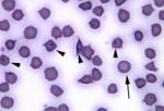
| − | + | [[Image:Theileria cervi.jpg|thumb|right|150px|''Theileria cervi'' (deer) - Drs. Elizabeth Howerth and Bruce LeRoy, Department of Pathology, UGA College of Veterinary Medicine]]# | |
| − | |||
| − | |||
| − | |||
| − | |||
| − | |||
| − | |||
| − | |||
| − | |||
| − | |||
| − | |||
| − | |||
| − | |||
| − | |||
| − | |||
| − | |||
| − | [[Image:Theileria parva life cycle.jpg|thumb|right| | ||
| − | [[ | ||
| − | |||
| − | ''''' | + | *Main species of veterinary importance is ''Theileria parva'' |
| + | **Causes '''East Coast Fever''' | ||
| + | ***Severe, proliferative lymphatic disease of cattle | ||
| + | ***Central and Eastern Africa | ||
| + | ***Transmitted by [[Hard Ticks - Overseas|''Rhipicephalus appendiculatus'']] | ||
| + | ***[[Ticks#Disease Transmission|Trans-stadial]] transmission | ||
| − | Other species cause | + | *Other ''Theileria'' species cause production losses in cattle and sheep in the Middle East, Mediterranean and in Northern Africa |
| − | + | '''Life Cycle''' | |
| − | + | *Incubation phase lasts 1 week | |
| − | |||
| − | |||
| − | + | *Lymphoblast proliferation | |
| + | **Local [[Lymph Nodes - Anatomy & Physiology|lymph node]] first infected then spreads through body | ||
| + | **Occurs in week two | ||
| − | + | *Lymphoid depletion | |
| + | **[[Lymphocytes - Introduction|Lymphocytes]] killed | ||
| + | **Decreases lymphopoiesis | ||
| + | **Occurs in week 3 | ||
| − | + | *Total incubation period takes about 18 days | |
| − | + | '''Diagnosis''' | |
| + | *Clinical signs | ||
| + | **Pyrexia | ||
| + | **Enlarged local [[Lymph Nodes - Anatomy & Physiology|lymph node]] | ||
| + | ***Usually parotid [[Lymph Nodes - Anatomy & Physiology|lymph node]] as [[Hard Ticks - Overseas|''Rhipicephalus appendiculatus'']] feeds in the ear | ||
| + | **Loss of condition | ||
| − | + | *Examine Giemsa stained smears of: | |
| + | **Local [[Lymph Nodes - Anatomy & Physiology|lymph node]] aspirated for schizonts | ||
| + | **Blood smears for piroplasms in red blood cells | ||
| − | + | *Post-mortem | |
| + | **Pulmonary oedema | ||
| + | **Gut mucosal haemorrhages | ||
| + | **[[Lymph Nodes - Anatomy & Physiology|Lymph node]] and [[Spleen - Anatomy & Physiology|splenic]] cellular atrophy | ||
| − | + | '''Control''' | |
| − | [[ | + | *Integrated control of both the [[Tick Control|tick vector]] and disease |
| + | **[[Vaccines|Vaccination]] and [[Ectoparasiticides]] | ||
| − | + | *Current [[Vaccines|vaccination]] is live unattentuated | |
| − | + | **Contains frozen stabilate of ground up tick gut containing infective sporozoites | |
| + | **Long lasting oxytetracycline administered at the same time to slow down schizogony giving the immune response time to develop | ||
| − | |||
| − | [[Babesiosis - Horse| | + | ''Theileria equi'' (formerly ''Babesia equi'') and ''Babesia caballi'' cause [[Babesiosis - Horse|babesiosis in horses]] |
| − | |||
| − | + | ==Test yourself with the Piroplasmida Flashcards== | |
| − | + | [[Piroplasmida_Flashcards|Piroplasmida Flashcards]] | |
| − | + | [[Category:Piroplasmida]] | |
| − | |||
| − | |||
| − | |||
| − | |||
| − | |||
| − | |||
| − | |||
| − | |||
| − | |||
| − | |||
| − | |||
| − | |||
| − | |||
| − | |||
| − | |||
| − | |||
| − | |||
| − | |||
| − | |||
| − | |||
| − | |||
| − | |||
| − | |||
| − | |||
| − | |||
| − | |||
| − | |||
| − | |||
| − | |||
| − | + | [[Category:To_Do_-_Parasites]] | |
| − | [[Category: | ||
| − | |||
| − | |||
Revision as of 12:01, 6 October 2010
File:Lymph node smear East Coast Fever.jpg
Lymph node smear of a cow with East Coast Fever - Drs. Elizabeth Howerth and Bruce LeRoy, Department of Pathology, UGA College of Veterinary Medicine
File:H and E stain brain East Coast Fever.jpg
H and E stain of brain and meningal vessels of a cow with East Coast Fever - Drs. Elizabeth Howerth and Bruce LeRoy, Department of Pathology, UGA College of Veterinary Medicine
#
- Main species of veterinary importance is Theileria parva
- Causes East Coast Fever
- Severe, proliferative lymphatic disease of cattle
- Central and Eastern Africa
- Transmitted by Rhipicephalus appendiculatus
- Trans-stadial transmission
- Causes East Coast Fever
- Other Theileria species cause production losses in cattle and sheep in the Middle East, Mediterranean and in Northern Africa
Life Cycle
- Incubation phase lasts 1 week
- Lymphoblast proliferation
- Local lymph node first infected then spreads through body
- Occurs in week two
- Lymphoid depletion
- Lymphocytes killed
- Decreases lymphopoiesis
- Occurs in week 3
- Total incubation period takes about 18 days
Diagnosis
- Clinical signs
- Pyrexia
- Enlarged local lymph node
- Usually parotid lymph node as Rhipicephalus appendiculatus feeds in the ear
- Loss of condition
- Examine Giemsa stained smears of:
- Local lymph node aspirated for schizonts
- Blood smears for piroplasms in red blood cells
- Post-mortem
- Pulmonary oedema
- Gut mucosal haemorrhages
- Lymph node and splenic cellular atrophy
Control
- Integrated control of both the tick vector and disease
- Current vaccination is live unattentuated
- Contains frozen stabilate of ground up tick gut containing infective sporozoites
- Long lasting oxytetracycline administered at the same time to slow down schizogony giving the immune response time to develop
Theileria equi (formerly Babesia equi) and Babesia caballi cause babesiosis in horses

