Difference between revisions of "Equine Internal Medicine Q&A 18"
Ggaitskell (talk | contribs) |
|||
| (2 intermediate revisions by the same user not shown) | |||
| Line 5: | Line 5: | ||
<br /> | <br /> | ||
| − | '''A 12-year-old pony gelding is presented with a three week history of weight loss, inappetence, intermittent fever (up to 38.7°C) and ventral thoracic and abdominal oedema. Auscultation of the chest reveals bilateral absence of ventral lung sounds, and a pleural effusion is confirmed by ultrasonography. Thoracocentesis yields a large volume of blood-stained watery fluid. A tracheal wash did not reveal | + | '''A 12-year-old pony gelding is presented with a three week history of weight loss, inappetence, intermittent fever (up to 38.7°C) and ventral thoracic and abdominal oedema. Auscultation of the chest reveals bilateral absence of ventral lung sounds, and a pleural effusion is confirmed by ultrasonography. Thoracocentesis yields a large volume of blood-stained watery fluid. A tracheal wash did not reveal any infectious cause.''' |
<br /> | <br /> | ||
| Line 13: | Line 13: | ||
|a1=Thoracic neoplasia. <br> | |a1=Thoracic neoplasia. <br> | ||
The commonest type of thoracic neoplasia in the horse is mediastinal lymphosarcoma. | The commonest type of thoracic neoplasia in the horse is mediastinal lymphosarcoma. | ||
| − | |l1= | + | |l1=Lymphoma |
|q2=How can the diagnosis be confirmed? | |q2=How can the diagnosis be confirmed? | ||
|a2= | |a2= | ||
| Line 21: | Line 21: | ||
*Mediastinal masses may sometimes extend through the thoracic inlet and be palpable externally at the base of the jugular groove; in this situation they may be amenable to biopsy. | *Mediastinal masses may sometimes extend through the thoracic inlet and be palpable externally at the base of the jugular groove; in this situation they may be amenable to biopsy. | ||
*In the case of lymphosarcoma, peripheral lymph nodes may be enlarged due to neoplastic infiltration, and these may be biopsied. | *In the case of lymphosarcoma, peripheral lymph nodes may be enlarged due to neoplastic infiltration, and these may be biopsied. | ||
| − | |l2= | + | |l2=Thoracocentesis#Equine Thoracocentesis |
</FlashCard> | </FlashCard> | ||
Latest revision as of 15:27, 26 February 2013
| This question was provided by Manson Publishing as part of the OVAL Project. See more Equine Internal Medicine questions |
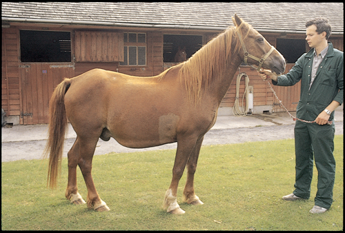
A 12-year-old pony gelding is presented with a three week history of weight loss, inappetence, intermittent fever (up to 38.7°C) and ventral thoracic and abdominal oedema. Auscultation of the chest reveals bilateral absence of ventral lung sounds, and a pleural effusion is confirmed by ultrasonography. Thoracocentesis yields a large volume of blood-stained watery fluid. A tracheal wash did not reveal any infectious cause.
| Question | Answer | Article | |
| What is the most likely diagnosis? | Thoracic neoplasia. The commonest type of thoracic neoplasia in the horse is mediastinal lymphosarcoma. |
Link to Article | |
| How can the diagnosis be confirmed? |
|
Link to Article | |
