Difference between revisions of "Small Animal Orthopaedics Q&A 04"
(Created page with "[[|centre|500px]] <br /> '''A photograph and craniocaudal view radiograph of the right distal antebrachium and carpus of a five-month-old, male Great Dane that is depressed, an...") |
|||
| (2 intermediate revisions by 2 users not shown) | |||
| Line 1: | Line 1: | ||
| − | + | {{Manson | |
| + | |book = Small Animal Orthopaedics Q&A}} | ||
| + | [[File:SmAnOrth 4a.jpg|centre|500px]] | ||
| + | <br> | ||
| + | [[File:SmAnOrth 4b.jpg|centre|500px]] | ||
| + | <br> | ||
| + | [[File:SmAnOrth 4c.jpg|centre|500px]] | ||
<br /> | <br /> | ||
| Line 8: | Line 14: | ||
<FlashCard questions="3"> | <FlashCard questions="3"> | ||
| − | |q1=Describe the radiographic | + | |q1=Describe the radiographic abnormalities and state a diagnosis. |
|a1= | |a1= | ||
The radiograph reveals an irregular radiolucent zone involving the metaphysis subjacent and parallel to the distal radial and ulnar physes. | The radiograph reveals an irregular radiolucent zone involving the metaphysis subjacent and parallel to the distal radial and ulnar physes. | ||
| Line 23: | Line 29: | ||
The bone scan of the forelimbs in this dog reveals increased up take in the regions where non-septic suppurative inflammation and trabecular infarction in the metaphyseal regions is present. | The bone scan of the forelimbs in this dog reveals increased up take in the regions where non-septic suppurative inflammation and trabecular infarction in the metaphyseal regions is present. | ||
| − | |l1= | + | |l1=Hypertrophic Osteodystrophy |
|q2=What is the etiology of this disease? | |q2=What is the etiology of this disease? | ||
|a2= | |a2= | ||
| Line 39: | Line 45: | ||
Although infectious agents have not been identified histologically or cultured from dogs with HOD, canine distemper virus has been | Although infectious agents have not been identified histologically or cultured from dogs with HOD, canine distemper virus has been | ||
detected in bone cells of dogs with HOD using in situ hybridization techniques. The association between HOD and canine distemper virus infection, however, remains speculative. | detected in bone cells of dogs with HOD using in situ hybridization techniques. The association between HOD and canine distemper virus infection, however, remains speculative. | ||
| − | |l2= | + | |l2=Hypertrophic Osteodystrophy |
|q3=What treatment is appropriate for this dog? | |q3=What treatment is appropriate for this dog? | ||
|a3= | |a3= | ||
| Line 51: | Line 57: | ||
Dogs that recover from HOD may have varying degrees of bone deformity depending on the severity of the disease. Permanent antebrachial bowing deformities associated with attenuated or arrested physeal growth can occur. | Dogs that recover from HOD may have varying degrees of bone deformity depending on the severity of the disease. Permanent antebrachial bowing deformities associated with attenuated or arrested physeal growth can occur. | ||
| − | |l3= | + | |l3=Hypertrophic Osteodystrophy#Treatment |
</FlashCard> | </FlashCard> | ||
Latest revision as of 23:14, 23 October 2011
| This question was provided by Manson Publishing as part of the OVAL Project. See more Small Animal Orthopaedics Q&A. |
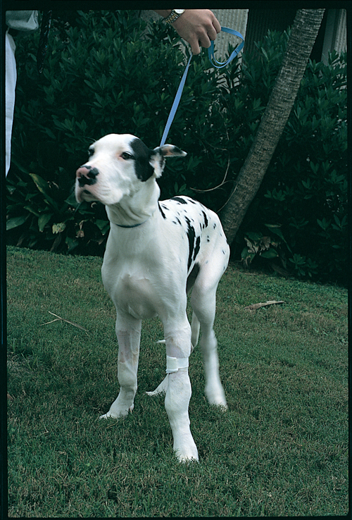
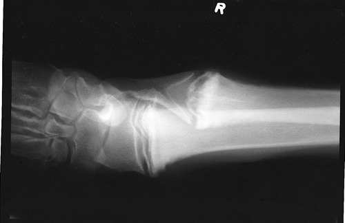
A photograph and craniocaudal view radiograph of the right distal antebrachium and carpus of a five-month-old, male Great Dane that is depressed, anorexic, febrile and reluctant to walk.
| Question | Answer | Article | |
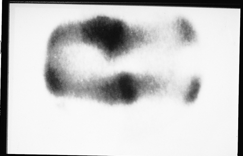
| Describe the radiographic abnormalities and state a diagnosis. | The radiograph reveals an irregular radiolucent zone involving the metaphysis subjacent and parallel to the distal radial and ulnar physes. These radiographic abnormalities are characteristic of early hypertrophic osteodystrophy (HOD). There is also a small retained cartilage core affecting the distal ulna. HOD is an acute suppurative inflammatory disease affecting long bones of rapidly growing, juvenile, large-breed dogs. The radiolucent areas seen radiographically correspond to regions of non-septic suppurative (predominantly non-degenerate neutrophils) inflammation. Metaphyseal trabecular infarction is seen and perimetaphysealperiosteal new bone is evident. The bone scan of the forelimbs in this dog reveals increased up take in the regions where non-septic suppurative inflammation and trabecular infarction in the metaphyseal regions is present. |
Link to Article | |
| What is the etiology of this disease? | The cause of HOD is unknown. The disease is not heritable. Hypovitaminosis C has been purported as a cause of HOD; however, dogs produce ascorbic acid endogenously and dogs with HOD have normal ascorbic acid levels. Supplementation of ascorbic acid neither resolves nor prevents recurrence of clinical abnormalities. Overnutrition with protein, vitamins such as vitamin D, minerals and energy have all been anecdotally associated with HOD, but experimental oversupplementation of these nutrients has not produced HOD. Correction of potential dietary influences does not alter the course of HOD. Although infectious agents have not been identified histologically or cultured from dogs with HOD, canine distemper virus has been detected in bone cells of dogs with HOD using in situ hybridization techniques. The association between HOD and canine distemper virus infection, however, remains speculative. |
Link to Article | |
| What treatment is appropriate for this dog? | Treatment of HOD is supportive and directed at maintenance of hydration, nutritional support and prevention of pressure sores. Exercise restriction is recommended to reduce the potential for additional bone infarction. Anti-inflammatory drugs can be prescribed to mitigate pain and pyrexia. Occasionally, a debilitated puppy will not respond and will die or be euthanized. Dogs that recover from HOD may have varying degrees of bone deformity depending on the severity of the disease. Permanent antebrachial bowing deformities associated with attenuated or arrested physeal growth can occur. |
Link to Article | |


