Hypertrophic Osteodystrophy
Also known as: Metaphyseal Osteopathy — HOD
Introduction
This is a developmental disease of young, large, rapidly-growing large and giant breeds such as the Great Dane, St Bernard, Irish Setter, Labrador, German Shepherd, Doberman and Weimaraner.
It most frequently presents at the age of 3-4 months (range 2-8) and males are affected more commonly than females.
The disease occurs at the metaphysis of long bones, especially the distal ulna and radius.
The aetiology of the condition is unknown, but theories include: an excessive plane of nutrition, calcium/phosphate imbalance, vitamin C deficiency or an infectious agent.
Clinical Signs
Respiratory or gastrointestinal illness may precede the onset of skeletal disease.
Clinical signs vary from mild lameness to severe systemic illness, pyrexia, depression, inappetance, weight loss and inability to stand.
Pain can be elicited by digital pressure on the metaphysis.
The condition is usually bilaterally symmetrical and may affect all four limbs.
In severe cases, the long bone metaphyses are visibly swollen, hot and painful on palpation.
Active periods of disease may last for several days and relapses may occur at intervals of 1-6 weeks.
Some severely suffering dogs may die or may be euthanised on owner's request.
Diagnosis
The clinical signs and presentation are suggestive.
Radiography can confirm the diagnosis. Findings include:
- irregular radiolucent line (double physeal line) in the metaphysis, parallel to the normal radiolucent physeal line
- the physis may appear widened
- subperiosteal new bone formation at the metaphysis forming a collar of bone, also known as bone cuffing
- evidence of growth deformities
Bone scintigraphy may reveal increased uptake of agent at the metaphysis.
Haematology and biochemistry are usually unremarkable.
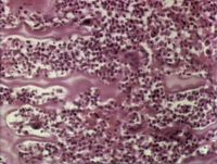
Histology of the area would reveal: haemorrhage and necrosis of osteoblasts in the growth plates and primary spongiosa, intense infiltration by neutrophils, periosteal reaction and formation of new bone on external surface above the lesion.
Treatment
HOD is usually self-limiting and mildly affected dogs recover within a few weeks.
Treatment involves supportive care with proper nutrition, fluids and analgesia. Buffered aspirin is the preferred analgesic.
In severe cases, corticosteroids and antibiotics may be indicated, depending on blood culture results. Recumbent puppies should be turned every 4 hours and placed in a well-padded cage.
Correction of angular limb deformities may be necessary.
Prognosis
This is related to disease severity.
Mildly affected dogs have a good prognosis and many recover spontaneously. Diaphyseal deformities can be severe but are usually not debilitating.
Severely affected dogs have a poor prognosis and some dogs can succumb to hyperthermia or acidosis. Euthanasia may also be performed on the worst cases.
| Hypertrophic Osteodystrophy Learning Resources | |
|---|---|
 Test your knowledge using flashcard type questions |
Small Animal Orthopaedics Q&A 04 |
References
Pasquini, C. (1999) Tschauner's Guide to Small Animal Clinics Sudz Publishing
Dunn, J. (1999) Textbook of small animal medicine Elsevier Health Sciences
Hosgood, G. (1998) Small animal paediatric medicine and surgery Elsevier Health Sciences
| This article has been peer reviewed but is awaiting expert review. If you would like to help with this, please see more information about expert reviewing. |
Error in widget FBRecommend: unable to write file /var/www/wikivet.net/extensions/Widgets/compiled_templates/wrt69a01e686a45a0_30187657 Error in widget google+: unable to write file /var/www/wikivet.net/extensions/Widgets/compiled_templates/wrt69a01e68736bf3_16996617 Error in widget TwitterTweet: unable to write file /var/www/wikivet.net/extensions/Widgets/compiled_templates/wrt69a01e687cb750_80571105
|
| WikiVet® Introduction - Help WikiVet - Report a Problem |
