Difference between revisions of "Pituitary Gland Flash Cards - Anatomy & Physiology"
Jump to navigation
Jump to search
m (New page: {{toplink |backcolour = FAFAD2 |linkpage =Endocrine System - Anatomy & Physiology |linktext =Endocrine System |maplink = Endocrine System (Content Map) - Anatomy & Physiology |pagetype =An...) |
m |
||
| (5 intermediate revisions by 2 users not shown) | |||
| Line 1: | Line 1: | ||
| − | + | ===Pituitary Gland=== | |
| − | + | <FlashCard questions="10"> | |
| − | + | |q1=Describe the location of the Pituitary Gland. | |
| − | + | |a1=The pituitary gland, or hypophysis lies within a bony cavity, the Sella Turcica, in the base of the skull just ventral to the hypothalamus. It lies between the more rostral Optic Chiasma, and the more caudal Mammillary Bodies. | |
| − | + | |l1=Pituitary Gland - Anatomy & Physiology | |
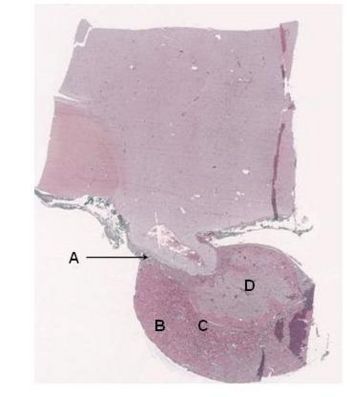
| − | + | |q2=Label the diagram ('Image 1' below). | |
| − | + | |a2= | |
| − | + | *A. Pars Tuberalis | |
| − | + | *B. Pars Distalis | |
| − | + | *C. Pars Intermedia | |
| − | + | *D. Pars Nervosa | |
| − | + | |l2=Pituitary Gland - Anatomy & Physiology | |
| − | + | |q3=Which sections of the Pituitary Gland labelled above make up the Adenohypophysis and Neurohypophysis: | |
| − | + | |a3= | |
| − | + | *Adenohypophysis (A) = Pars Tuberalis, B = Pars Distalis C = Pars IntermediaB,C | |
| − | ==Pituitary Gland== | + | *Neurohypophysis (D) = Pars Nervosa |
| − | + | |l3=Pituitary Gland - Anatomy & Physiology | |
| − | + | |q4=What is the blood supply to the Pituitary Gland: | |
| − | + | |a4=Superior Hypophyseal Arteries | |
| − | + | |l4=Pituitary Gland - Anatomy & Physiology | |
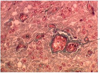
| − | + | |q5=What part of the Pituitary Gland does this histological section ('Image 2' below) represent? | |
| − | + | |a5=Pars Nervosa | |
| − | + | |l5=Pituitary Gland - Anatomy & Physiology#Histology Gallery | |
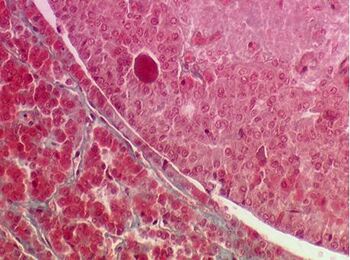
| − | + | |q6=Which parts of the Pituitary Gland does this histological section ('Image 3' below) represent? | |
| − | + | |a6= | |
| − | | | ||
| − | |||
| − | | | ||
| − | | | ||
| − | |||
| − | |||
| − | | | ||
| − | *A | ||
| − | *B | ||
| − | *C | ||
| − | *D | ||
| − | | | ||
| − | | | ||
| − | |||
| − | | | ||
| − | *Adenohypophysis | ||
| − | *Neurohypophysis | ||
| − | | | ||
| − | | | ||
| − | |||
| − | | | ||
| − | |||
| − | | | ||
| − | | | ||
| − | |||
| − | |||
| − | | | ||
| − | |||
| − | | | ||
| − | | | ||
| − | |||
| − | |||
| − | | | ||
*Pars Intermedia (Upper Right) | *Pars Intermedia (Upper Right) | ||
*Pars Distalis (Lower Left) | *Pars Distalis (Lower Left) | ||
*Sections Separated by the residual lumen of Rathke's Pouch - the Hypophyseal Cleft. | *Sections Separated by the residual lumen of Rathke's Pouch - the Hypophyseal Cleft. | ||
| − | | | + | |l6=Pituitary Gland - Anatomy & Physiology#Histology Gallery |
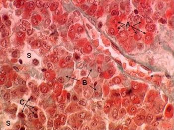
| − | | | + | |q7=What part of the Pituitary Gland does this histological section ('Image 4' below) represent? |
| − | + | |a7=Pars Distalis | |
| − | + | |l7=Pituitary Gland - Anatomy & Physiology#Histology Gallery | |
| − | + | |q8=What are the cell types within the Pars Distalis and what hormones do they produce? | |
| − | + | |a8= | |
| − | | | ||
| − | | | ||
| − | |||
| − | | | ||
*Corticotropes - POMC which is then cleaved to Adrenocorticotropin Hormone - ACTH | *Corticotropes - POMC which is then cleaved to Adrenocorticotropin Hormone - ACTH | ||
*Thyrotropes - Thyroid Stimulating Hormone | *Thyrotropes - Thyroid Stimulating Hormone | ||
| Line 75: | Line 38: | ||
*Lactotropes - Prolactin | *Lactotropes - Prolactin | ||
*Somatotropes - Growth Hormone. | *Somatotropes - Growth Hormone. | ||
| − | | | + | |l8=Pituitary Gland - Anatomy & Physiology#Hormones of the Anterior Pituitary Gland |
| − | | | + | |q9=What is secreted by the Posterior Pituitary Gland and what are their actions: |
| − | + | |a9= | |
| − | | | ||
*Oxytocin - Promotes milk let down and uterine contractions during parturtion. | *Oxytocin - Promotes milk let down and uterine contractions during parturtion. | ||
*Anti Diuretic Hormone - ADH - Acts on the renal tubules to minimise water loss into the urine. | *Anti Diuretic Hormone - ADH - Acts on the renal tubules to minimise water loss into the urine. | ||
| − | | | + | |l9=Pituitary Gland - Anatomy & Physiology#Posterior Pituitary Gland |
| − | | | + | |q10=Decribe how Milk Let-Down is initiated. |
| − | + | |a10= | |
| − | | | ||
*Impulses travel via superfical sensory pathways and the inguinal nerve. | *Impulses travel via superfical sensory pathways and the inguinal nerve. | ||
*Afferent sensory neurons enter the lumbar part of the spinal cord to the thalmus. | *Afferent sensory neurons enter the lumbar part of the spinal cord to the thalmus. | ||
| Line 93: | Line 54: | ||
*Resistance in excretory ducts and teat canal is reduced. | *Resistance in excretory ducts and teat canal is reduced. | ||
*Increased milk outflow. | *Increased milk outflow. | ||
| − | | | + | |l10=Pituitary Gland - Anatomy & Physiology#Oxytocin |
| − | | | + | </FlashCard> |
| + | |||
| + | {| | ||
| + | !Image 1 | ||
| + | |[[Image:PituitaryGlandFlashCard1.jpg|350px|©RVC 2008]] | ||
| + | |- | ||
| + | !Image 2 | ||
| + | |[[Image:PituitaryGlandFlashCard2.jpg|350px|©RVC 2008]] | ||
| + | |- | ||
| + | !Image 3 | ||
| + | |[[Image:PituitaryGlandFlashCard3.jpg|350px|©RVC 2008]] | ||
| + | |- | ||
| + | !Image 4 | ||
| + | |[[Image:PituitaryGlandFlashCard4.jpg|350px|©RVC 2008]] | ||
| + | |] | ||
| + | |||
| + | |||
| + | [[Category:Endocrine System - Anatomy & Physiology]] | ||
| + | [[Category:Endocrine System Anatomy & Physiology Flashcards]] | ||
| + | [[Category:Image Review]] | ||
Latest revision as of 12:21, 21 June 2011
Pituitary Gland
| Question | Answer | Article | |
| Describe the location of the Pituitary Gland. | The pituitary gland, or hypophysis lies within a bony cavity, the Sella Turcica, in the base of the skull just ventral to the hypothalamus. It lies between the more rostral Optic Chiasma, and the more caudal Mammillary Bodies.
|
Link to Article | |
| Label the diagram ('Image 1' below). |
|
Link to Article | |
| Which sections of the Pituitary Gland labelled above make up the Adenohypophysis and Neurohypophysis: |
|
Link to Article | |
| What is the blood supply to the Pituitary Gland: | Superior Hypophyseal Arteries
|
Link to Article | |
| What part of the Pituitary Gland does this histological section ('Image 2' below) represent? | Pars Nervosa
|
Link to Article | |
| Which parts of the Pituitary Gland does this histological section ('Image 3' below) represent? |
|
Link to Article | |
| What part of the Pituitary Gland does this histological section ('Image 4' below) represent? | Pars Distalis
|
Link to Article | |
| What are the cell types within the Pars Distalis and what hormones do they produce? |
|
Link to Article | |
| What is secreted by the Posterior Pituitary Gland and what are their actions: |
|
Link to Article | |
| Decribe how Milk Let-Down is initiated. |
|
Link to Article | |
| Image 1 | 
| |
|---|---|---|
| Image 2 | 
| |
| Image 3 | 
| |
| Image 4 | 
|
] |