Difference between revisions of "Pelvis - Anatomy & Physiology"
Fiorecastro (talk | contribs) |
|||
| (10 intermediate revisions by 4 users not shown) | |||
| Line 1: | Line 1: | ||
| − | {{ | + | {{OpenPagesTop}} |
| − | |||
| − | |||
| − | |||
| − | |||
| − | |||
| − | }} | ||
| − | |||
==Pelvic Girdle== | ==Pelvic Girdle== | ||
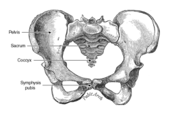
| − | [[Image:Pelvis.png|thumb|right| | + | [[Image:Pelvis.png|thumb|right|250px|Pelvis - Wikimedia Commons 2008]] |
| − | + | ||
| − | + | The pelvic girdle consists of two symmetrical halves. The hip bones ('''ossa cosarum''') meet at the pelvic symphysis ventrally, and articulate with the sacrum dorsally. | |
| − | + | ||
| − | + | ===Hip Bones=== | |
| − | + | ||
| − | + | Three bones develop from separate ossifications, within a single cartilage plate. | |
| − | + | ||
| − | + | 1. [[Hindlimb - Anatomy & Physiology#Ilium|'''Ilium''']] | |
| − | + | ||
| − | + | The ilium is craniodorsal and extends obliquely forward from the hip to articulate with the sacrum. The cranial wing varies between species. Dorsally, it forms the '''sacral tuber''', which is more prominent in larger animals than in dogs and cats. Ventrally, it forms the '''tuber coxae''', or the point of the hip. The margin of the wing is known as the '''iliac crest''' and the body is deeply excavated for attachment of the gluteus medius. The '''greater sciatic notch''' on dorsal border of the wing, is cut away at its junction with the shaft, to allow the sciatic nerve passage en route to the hind limb. | |
| − | + | ||
| − | + | 2. [[Hindlimb - Anatomy & Physiology#Pubis|'''Pubis''']] | |
| − | + | ||
| − | + | The pubis extends medially from the joint to form the cranial pelvic floor. It is L-shaped to give two branches; cranial (acetabular) and caudal (symphysial). | |
| − | + | ||
| − | + | 3. [[Hindlimb - Anatomy & Physiology#Ischium|'''Ischium''']] | |
| − | + | ||
| − | + | The ischium is caudal and forms most of the pelvic floor. The '''ischial tuberosity''' is formed by the caudolateral corner of the horizontal plate of the ischium. The '''pelvic symphysis''' comprises both the pubis and the ischium . | |
| − | + | ||
| + | The '''Acetabulum''' provides the socket to the joint of the hip, and is composed of all three bones of the pelvis. | ||
==Pelvic Joints and Ligaments== | ==Pelvic Joints and Ligaments== | ||
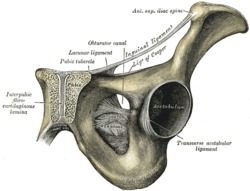
| − | [[Image:Grays Pelvic Symphysis.png|thumb|right| | + | [[Image:Grays Pelvic Symphysis.png|thumb|right|250px|Pelvic Symphysis, Gray's Anatomy - Wikimedia Commons 2008]] |
| − | + | ||
| − | + | '''Pelvic Symphysis''': secondary cartilaginous joint that ossifies with age and may expand in parturition. | |
| − | + | ||
| − | + | '''Sacroiliac joints''': synovial joints combined with fibrous joints. They transmit the weight of the trunk to the hindlimbs. | |
| − | + | ||
| − | + | '''Sacrotuberous ligament''': varies tremendously between species, the caudal edge is palpable. | |
| − | + | ||
| − | + | ==Species differences== | |
| + | |||
| + | Larger species have a more vertical ilium, bringing the sacroiliac joint (and with it the weight of the trunk) closer to the hip. Smaller species have an oblique ilium. The dog has a stout cord extending between the sacrum and lateral ischial tuberosity, this is not present in the cat. In ungulates, the '''sacrosciatic ligament''' expands to a broad sheet. | ||
| + | |||
| + | {{Template:Learning | ||
| + | |videos = [http://stream2.rvc.ac.uk/Frean/sheep/SacroSciaticLigament.wmv Sacrosciatic Ligament of the Sheep] | ||
| + | |CAL = [[Canine Radiographic Anatomy resource|Interactive programme where you can explore the radiographic anatomy of the dog, including the pelvis]] | ||
| + | |dragster = [[Canine Pelvis Radiographical Anatomy Resources (I & II)]]<br>[[Canine Pelvis Skeletal Anatomy Resources (I & II)]]<br>[[Canine Pelvis Surface Anatomy Resources (I & II)]]<br>[[Canine Pelvis and Femoral Region Surface Anatomy Resource]]<br>[[Equine Abdomen and Hip Surface Anatomy Resource]]<br>[[Equine Pelvis and Genitalia Surface Anatomy Resource]] | ||
| + | |OVAM = [http://www.onlineveterinaryanatomy.net/content/muscle-flashcards-hip-quicktime Muscle flashcards - muscles of the canine hip] | ||
| + | }} | ||
| + | |||
| + | |||
| + | ==Webinars== | ||
| + | <rss max="10" highlight="pelvis">https://www.thewebinarvet.com/orthopaedics/webinars/feed</rss> | ||
| + | |||
| + | [[Category:Musculoskeletal System - Anatomy & Physiology]] | ||
| + | [[Category:A&P Done]] | ||
Latest revision as of 21:27, 28 November 2022
Pelvic Girdle
The pelvic girdle consists of two symmetrical halves. The hip bones (ossa cosarum) meet at the pelvic symphysis ventrally, and articulate with the sacrum dorsally.
Hip Bones
Three bones develop from separate ossifications, within a single cartilage plate.
1. Ilium
The ilium is craniodorsal and extends obliquely forward from the hip to articulate with the sacrum. The cranial wing varies between species. Dorsally, it forms the sacral tuber, which is more prominent in larger animals than in dogs and cats. Ventrally, it forms the tuber coxae, or the point of the hip. The margin of the wing is known as the iliac crest and the body is deeply excavated for attachment of the gluteus medius. The greater sciatic notch on dorsal border of the wing, is cut away at its junction with the shaft, to allow the sciatic nerve passage en route to the hind limb.
2. Pubis
The pubis extends medially from the joint to form the cranial pelvic floor. It is L-shaped to give two branches; cranial (acetabular) and caudal (symphysial).
3. Ischium
The ischium is caudal and forms most of the pelvic floor. The ischial tuberosity is formed by the caudolateral corner of the horizontal plate of the ischium. The pelvic symphysis comprises both the pubis and the ischium .
The Acetabulum provides the socket to the joint of the hip, and is composed of all three bones of the pelvis.
Pelvic Joints and Ligaments
Pelvic Symphysis: secondary cartilaginous joint that ossifies with age and may expand in parturition.
Sacroiliac joints: synovial joints combined with fibrous joints. They transmit the weight of the trunk to the hindlimbs.
Sacrotuberous ligament: varies tremendously between species, the caudal edge is palpable.
Species differences
Larger species have a more vertical ilium, bringing the sacroiliac joint (and with it the weight of the trunk) closer to the hip. Smaller species have an oblique ilium. The dog has a stout cord extending between the sacrum and lateral ischial tuberosity, this is not present in the cat. In ungulates, the sacrosciatic ligament expands to a broad sheet.
| Pelvis - Anatomy & Physiology Learning Resources | |
|---|---|
 Test your knowledge using drag and drop boxes |
Canine Pelvis Radiographical Anatomy Resources (I & II) Canine Pelvis Skeletal Anatomy Resources (I & II) Canine Pelvis Surface Anatomy Resources (I & II) Canine Pelvis and Femoral Region Surface Anatomy Resource Equine Abdomen and Hip Surface Anatomy Resource Equine Pelvis and Genitalia Surface Anatomy Resource |
 Selection of relevant videos |
Sacrosciatic Ligament of the Sheep |
 Selection of relevant programmes to enhance your knowledge |
Interactive programme where you can explore the radiographic anatomy of the dog, including the pelvis |
Anatomy Museum Resources |
Muscle flashcards - muscles of the canine hip |
Webinars
Failed to load RSS feed from https://www.thewebinarvet.com/orthopaedics/webinars/feed: Error parsing XML for RSS

