Difference between revisions of "Coccidiosis - Horse"
Fiorecastro (talk | contribs) |
|||
| (17 intermediate revisions by 3 users not shown) | |||
| Line 1: | Line 1: | ||
| + | {{OpenPagesTop}} | ||
| + | ==Introduction== | ||
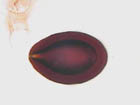
[[Image:Eimeria leukarti horse.jpg|thumb|right|150px|''Eimeria leukarti'' - Joaquim Castellà Veterinary Parasitology Universitat Autònoma de Barcelona]] | [[Image:Eimeria leukarti horse.jpg|thumb|right|150px|''Eimeria leukarti'' - Joaquim Castellà Veterinary Parasitology Universitat Autònoma de Barcelona]] | ||
| − | + | Equine coccidiosis is an infection of the intestinal tract of equids by [[:Category:Protozoa|protozoa]] of the genera [[Eimeria spp.|''Eimeria'']]. Horses may be infected by 3 species of ''Eimeria''; ''Eimeria leukarti'', ''E. solipedum'' and ''E. uniungulsti''. The most common coccidial oocyst identified in equine faeces is that of ''E. leukarti''. The prepatent period of infection by the protozoa is 16-35 days. Infection follows the ingestion of sporulated oocysts in contaminated food or water. After exposure to bile in the small intestine, the oocysts excyst and individual sporozoites emerge. Oocysts, released as the final product of the sexual reproductive cycle are shed in the faeces and are infective to new hosts. [[:Category:Coccidia|Coccidia]] are a common incidental finding in normal foals aged 30-125 days and suggest that this organism does not cause clinical signs in foals. | |
| − | + | ==Clinical signs== | |
| + | Clinical signs associated with the condition occur due to the damaged intestinal epithelium and connective tissue underlying the mucosa. These may include profuse diarrhoea, weight loss, ventral oedema, intestinal haemorrhage and catarrhal inflammation of the intestinal tract. | ||
| − | + | ==Diagnosis== | |
| − | + | A diagnosis of coccidiosis is made by detection of oocysts in the faeces using faecal flotation. A solution of saturated sugar or sodium nitrate solution is used. Oocysts are approximately 12μm in diameter and are identified by their dark brown colour and thick-walled, ovoid shape. They also contain a prominent micropyle on the narrower end. | |
| − | |||
| − | *'' | + | ==Treatment== |
| − | [[Category: | + | The treatment of choice for the disease is the sulphonamide antimicrobial agents. Sulphamethazine or Sulphathiazole are commonly used. Prevention of coccidiosis is obtained by providing adequate nutrition and hygiene and minimising stress. |
| + | |||
| + | {{Learning | ||
| + | |literature search = [http://www.cabdirect.org/search.html?q=(title:(Coccidiosis)+OR+title:(coccidia))+AND+od:(horses) Coccidiosis in horses publications] | ||
| + | }} | ||
| + | |||
| + | ==References== | ||
| + | *Bertone, J., Horspool, L. J. I. (2004) '''Equine Clinical Pharmacology''' ''Elsevier Health Sciences'' | ||
| + | *Lavoie, J. P., Hinchcliff, K. W. (2009) '''Blackwell's Five-Minute Veterinary Consult: Equine''' ''John Wiley and Sons'' | ||
| + | *Sellon, D. C., Long, M. T. (2007) '''Equine Infectious Diseases''' ''Elsevier Health Sciences'' | ||
| + | |||
| + | |||
| + | {{review}} | ||
| + | ==Webinars== | ||
| + | <rss max="10" highlight="none">https://www.thewebinarvet.com/parasitology/webinars/feed</rss> | ||
| + | |||
| + | [[Category:Expert Review]] | ||
| + | [[Category:Respiratory Parasitic Infections]] | ||
| + | [[Category:Respiratory Diseases - Horse]][[Category:Alimentary Diseases - Horse]] | ||
Latest revision as of 14:42, 25 November 2022
Introduction
Equine coccidiosis is an infection of the intestinal tract of equids by protozoa of the genera Eimeria. Horses may be infected by 3 species of Eimeria; Eimeria leukarti, E. solipedum and E. uniungulsti. The most common coccidial oocyst identified in equine faeces is that of E. leukarti. The prepatent period of infection by the protozoa is 16-35 days. Infection follows the ingestion of sporulated oocysts in contaminated food or water. After exposure to bile in the small intestine, the oocysts excyst and individual sporozoites emerge. Oocysts, released as the final product of the sexual reproductive cycle are shed in the faeces and are infective to new hosts. Coccidia are a common incidental finding in normal foals aged 30-125 days and suggest that this organism does not cause clinical signs in foals.
Clinical signs
Clinical signs associated with the condition occur due to the damaged intestinal epithelium and connective tissue underlying the mucosa. These may include profuse diarrhoea, weight loss, ventral oedema, intestinal haemorrhage and catarrhal inflammation of the intestinal tract.
Diagnosis
A diagnosis of coccidiosis is made by detection of oocysts in the faeces using faecal flotation. A solution of saturated sugar or sodium nitrate solution is used. Oocysts are approximately 12μm in diameter and are identified by their dark brown colour and thick-walled, ovoid shape. They also contain a prominent micropyle on the narrower end.
Treatment
The treatment of choice for the disease is the sulphonamide antimicrobial agents. Sulphamethazine or Sulphathiazole are commonly used. Prevention of coccidiosis is obtained by providing adequate nutrition and hygiene and minimising stress.
| Coccidiosis - Horse Learning Resources | |
|---|---|
 Search for recent publications via CAB Abstract (CABI log in required) |
Coccidiosis in horses publications |
References
- Bertone, J., Horspool, L. J. I. (2004) Equine Clinical Pharmacology Elsevier Health Sciences
- Lavoie, J. P., Hinchcliff, K. W. (2009) Blackwell's Five-Minute Veterinary Consult: Equine John Wiley and Sons
- Sellon, D. C., Long, M. T. (2007) Equine Infectious Diseases Elsevier Health Sciences
| This article has been peer reviewed but is awaiting expert review. If you would like to help with this, please see more information about expert reviewing. |
Webinars
Failed to load RSS feed from https://www.thewebinarvet.com/parasitology/webinars/feed: Error parsing XML for RSS
