Difference between revisions of "Rectum - Anatomy & Physiology"
(→Links) |
Fiorecastro (talk | contribs) |
||
| (23 intermediate revisions by 4 users not shown) | |||
| Line 1: | Line 1: | ||
| − | {{ | + | {{OpenPagesTop}} |
| − | |||
| − | |||
| − | |||
| − | |||
| − | |||
| − | |||
| − | |||
| − | }} | ||
| − | |||
==Introduction== | ==Introduction== | ||
| − | [[Image:Puppy rectum-anus.JPG|thumb|right| | + | [[Image:Puppy rectum-anus.JPG|thumb|right|250px|Section of puppy showing rectum and anus copyright RVC 2008]] |
| + | |||
The rectum lies between the terminal portion of the descending [[Colon - Anatomy & Physiology|colon]] and [[Anus - Anatomy & Physiology|anus]]. It is empty most of the time, except after the mass movements of the [[Large Intestine - Anatomy & Physiology|large intestine]] which move faeces into the rectum. This stimulates defeaction, which may happen when an animal is frightened. | The rectum lies between the terminal portion of the descending [[Colon - Anatomy & Physiology|colon]] and [[Anus - Anatomy & Physiology|anus]]. It is empty most of the time, except after the mass movements of the [[Large Intestine - Anatomy & Physiology|large intestine]] which move faeces into the rectum. This stimulates defeaction, which may happen when an animal is frightened. | ||
==Structure== | ==Structure== | ||
| − | + | The rectum exists dorsal to the [[Reproductive System Overview - Anatomy & Physiology|reproductive organs]], [[Urinary System Overview - Anatomy & Physiology|bladder]] and [[Urethra - Anatomy & Physiology|urethra]]. The cranial portion of the rectum is attached to the dorsal body wall by a short mesorectum which is a continuation of the mesocolon. The mesorectum is reflected to continue with the parietal [[Peritoneal Cavity - Anatomy & Physiology|peritoneum]] of the pelvic cavity and to cover the [[Urinary System Overview - Anatomy & Physiology|urogenital organs]] ventrally. This forms the '''rectogenital pouch''', therefore the most distal part of the rectum is retroperitoneal. This distal, retroperitoneal part is directly attached to the [[Vagina and Vestibule - Anatomy & Physiology|vagina]] in the female and to the [[Urethra - Anatomy & Physiology|urethra]] in the male. The retroperitoneal space is filled with soft tissue rich in fat. | |
| − | |||
| − | |||
| − | |||
| − | |||
| − | |||
| − | |||
==Function== | ==Function== | ||
'''Defeacation''' | '''Defeacation''' | ||
| − | |||
| − | |||
| − | |||
| − | |||
| − | |||
| − | |||
| − | |||
| − | |||
| − | |||
| − | |||
| − | |||
| − | |||
| − | + | After the mass movements of the [[Large Intestine - Anatomy & Physiology|large intestine]], the rectum becomes filled with faeces. This stimulates pressure sensitive cells in the wall of the rectum and initiates the defeacation reflex. The reflex causes a forceful contraction of the rectum, and relaxation of the internal anal sphincter. This produces the conscious sensation of the need to empty the bowel. Some species (see [[#Species Differences|species differences]]) are able to voluntarily keep the external anal sphincter closed if defeacation is not suitable in the situation. This reduces the defeaction reflex and reduces the conscious perception of needing to empty the bowel, until another mass movement occurs and a fresh reflex is created. | |
| − | [[ | ||
| − | |||
| − | |||
| − | |||
| − | |||
==Species Differences== | ==Species Differences== | ||
===Carnivore=== | ===Carnivore=== | ||
| − | + | ||
| − | + | Carnivore's control over the external anal sphincter is learned early in life. The defeaction reflex is increased by contraction of the abdominal muscles and closure of the vocal cords to increase the pressure in the abdominal cavity. | |
===Ruminant=== | ===Ruminant=== | ||
| − | |||
| − | + | Ruminants appear to lack the ability to control the external anal sphincter. | |
| − | |||
| − | == | + | ===Equine=== |
| − | + | Equine species appear to lack the ability to control the external anal sphincter. | |
| + | |||
| + | ==Histology== | ||
| + | [[Image:Recto-Anal Junction.jpg|thumb|right|250px|Recto-Anal Junction, from the [[Gastrointestinal Tract Histology resource|Gastrointestinal tract part 1 PowerPoint]] ]] | ||
| + | The epithelium of the rectum is '''columnar'''. '''Goblet cells''' are present in the mucosa. | ||
| + | |||
| + | ===Recto-Anal Junction=== | ||
| + | |||
| + | The junction marks the termination of the '''lamina muscularis''' and longitudinal layer of the '''tunica muscularis'''. The circular layer of the '''tunica muscularis''' forms the '''internal anal sphincter'''. The '''external anal sphincter''' is formed from skeletal muscle. At the junction, the epithelium changes from columnar to stratified squamous non-keratinised. | ||
==Links== | ==Links== | ||
| − | + | '''Click here for information on [[Intestines, Small and Large - Pathology|Pathology Of The Small and Large Intestines]]''' | |
| + | |||
| + | {{Template:Learning | ||
| + | |flashcards = [[Rectum - Anatomy & Physiology - Flashcards|Anatomy of the Rectum]] | ||
| + | |powerpoints = [[Gastrointestinal Tract Histology resource|Histology of the Gastrointestinal tract, see part 1]] | ||
| + | }} | ||
| + | |||
| + | ==Webinars== | ||
| + | <rss max="10" highlight="none">https://www.thewebinarvet.com/clinical-anatomy/webinars/feed</rss> | ||
| + | [[Category:Large Intestine - Anatomy & Physiology]] | ||
| + | [[Category:A&P Done]] | ||
Latest revision as of 21:16, 28 November 2022
Introduction
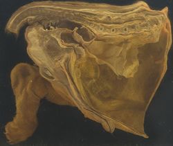
The rectum lies between the terminal portion of the descending colon and anus. It is empty most of the time, except after the mass movements of the large intestine which move faeces into the rectum. This stimulates defeaction, which may happen when an animal is frightened.
Structure
The rectum exists dorsal to the reproductive organs, bladder and urethra. The cranial portion of the rectum is attached to the dorsal body wall by a short mesorectum which is a continuation of the mesocolon. The mesorectum is reflected to continue with the parietal peritoneum of the pelvic cavity and to cover the urogenital organs ventrally. This forms the rectogenital pouch, therefore the most distal part of the rectum is retroperitoneal. This distal, retroperitoneal part is directly attached to the vagina in the female and to the urethra in the male. The retroperitoneal space is filled with soft tissue rich in fat.
Function
Defeacation
After the mass movements of the large intestine, the rectum becomes filled with faeces. This stimulates pressure sensitive cells in the wall of the rectum and initiates the defeacation reflex. The reflex causes a forceful contraction of the rectum, and relaxation of the internal anal sphincter. This produces the conscious sensation of the need to empty the bowel. Some species (see species differences) are able to voluntarily keep the external anal sphincter closed if defeacation is not suitable in the situation. This reduces the defeaction reflex and reduces the conscious perception of needing to empty the bowel, until another mass movement occurs and a fresh reflex is created.
Species Differences
Carnivore
Carnivore's control over the external anal sphincter is learned early in life. The defeaction reflex is increased by contraction of the abdominal muscles and closure of the vocal cords to increase the pressure in the abdominal cavity.
Ruminant
Ruminants appear to lack the ability to control the external anal sphincter.
Equine
Equine species appear to lack the ability to control the external anal sphincter.
Histology

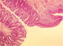
The epithelium of the rectum is columnar. Goblet cells are present in the mucosa.
Recto-Anal Junction
The junction marks the termination of the lamina muscularis and longitudinal layer of the tunica muscularis. The circular layer of the tunica muscularis forms the internal anal sphincter. The external anal sphincter is formed from skeletal muscle. At the junction, the epithelium changes from columnar to stratified squamous non-keratinised.
Links
Click here for information on Pathology Of The Small and Large Intestines
| Rectum - Anatomy & Physiology Learning Resources | |
|---|---|
 Test your knowledge using flashcard type questions |
Anatomy of the Rectum |
 Selection of relevant PowerPoint tutorials |
Histology of the Gastrointestinal tract, see part 1 |
Webinars
Failed to load RSS feed from https://www.thewebinarvet.com/clinical-anatomy/webinars/feed: Error parsing XML for RSS
