Difference between revisions of "Lizard and Snake Imaging"
| (5 intermediate revisions by the same user not shown) | |||
| Line 1: | Line 1: | ||
| − | {{ | + | {{OpenPagesTop}} |
[[Image:Main screen.jpg|200px|thumb|right|(Source RVC)]] | [[Image:Main screen.jpg|200px|thumb|right|(Source RVC)]] | ||
[[Image:Xray_liz.jpg|150px|thumb|right|(Source RVC)]] | [[Image:Xray_liz.jpg|150px|thumb|right|(Source RVC)]] | ||
| Line 32: | Line 32: | ||
Specific knowledge of the normal anatomy of individual species is important. The absence of an epiglottis facilitates tracheoscopy but the glottis is capable of spasm and can be easily damaged. The air sac and extensive rib cage of snakes means that insufflation is not always necessary. The anatomy of the thin, sac-like, lung means that it can be easily punctured. The long, relatively straight gastrointestinal tract facilitates examination. The vent may permit access to the colon and urinary and reproductive openings. | Specific knowledge of the normal anatomy of individual species is important. The absence of an epiglottis facilitates tracheoscopy but the glottis is capable of spasm and can be easily damaged. The air sac and extensive rib cage of snakes means that insufflation is not always necessary. The anatomy of the thin, sac-like, lung means that it can be easily punctured. The long, relatively straight gastrointestinal tract facilitates examination. The vent may permit access to the colon and urinary and reproductive openings. | ||
| − | == | + | {{Learning |
| − | [[ | + | |literature search = [http://www.cabdirect.org/search.html?start=0&q=((((subject:(radiography)+OR+subject:(endoscopy)+OR+subject:(coelioscopy)+OR+subject:(ultrasonography)+OR+subject:(ultrasound)+OR+subject:(ECG)+OR+subject:(echocardiography)))+OR+((title:(radiography)+OR+title:(endoscopy)+OR+title:(coelioscopy)+OR+title:(ultrasonography)+OR+title:(ultrasound)+OR+title:(ECG)+OR+title:(echocardiography)))))+AND+((((title:(lizard)+OR+ab:(lizard)+OR+od:(lizards)))+OR+((title:(snake)+OR+ab:(snake)+OR+od:(snakes))))) Lizard and Snake Imaging publications] |
| + | |full text = [http://www.cabi.org/cabdirect/FullTextPDF/2009/20093018983.pdf ''' Snake radiology: the essentials!''' Hernandez-Divers, S. J.; The North American Veterinary Conference, Gainesville, USA, Small animal and exotics. Proceedings of the North American Veterinary Conference, Volume 22, Orlando, Florida, USA, 2008, 2008, pp 1772-1774, 3 ref.] | ||
| + | |||
| + | [http://www.cabi.org/cabdirect/FullTextPDF/2007/20073120027.pdf '''Diagnostic imaging of lizards.''' Mitchell, M. A.; The North American Veterinary Conference, Gainesville, USA, Small animal and exotics. Proceedings of the North American Veterinary Conference, Volume 21, Orlando, Florida, USA, 2007, 2007, pp 1590-1591] | ||
| + | |||
| + | [http://www.cabi.org/cabdirect/FullTextPDF/2006/20063121826.pdf '''Reptile radiology: techniques, tips and pathology.''' Hernandez-Divers, S. J.; The North American Veterinary Conference, Gainesville, USA, Small animal and exotics. Proceedings of the North American Veterinary Conference, Volume 20, Orlando, Florida, USA, 7-11 January, 2006, 2006, pp 1626-1630, 4 ref.] | ||
| + | |||
| + | [http://www.cabi.org/cabdirect/FullTextPDF/2010/20103181633.pdf '''Rigid endoscopy in reptiles: not just for the coelom anymore - an introduction to pneumonoscopy and cloacoscopy.''' Stahl, S. J.; The North American Veterinary Conference, Gainesville, USA, Small animal and exotics. Proceedings of the North American Veterinary Conference, Orlando, Florida, USA, 16-20 January 2010, 2010, pp 1720-1721, 12 ref.] | ||
| + | |||
| + | [http://www.cabi.org/cabdirect/FullTextPDF/2009/20093118400.pdf ''' Is that normal? Clinical cases and standardization of the two-dimensional echocardiographic examination in snakes.''' Schilliger, L.; The North American Veterinary Conference, Gainesville, USA, Small animal and exotics. Proceedings of the North American Veterinary Conference, Orlando, Florida, USA, 17-21 January, 2009, 2009, pp 1807-1809] | ||
| + | [http://www.cabi.org/cabdirect/FullTextPDF/2009/20093118445.pdf ''' Fish, frogs, birds, elephants, and more: laparoscopy of nondomestic species.''' Stetter, M.; The North American Veterinary Conference, Gainesville, USA, Small animal and exotics. Proceedings of the North American Veterinary Conference, Orlando, Florida, USA, 17-21 January, 2009, 2009, pp 1940-1941, 12 ref.] | ||
| − | + | [http://www.cabi.org/cabdirect/FullTextPDF/2009/20093118399.pdf '''From A to Z: how to perform a diagnostic ultrasound examination in reptiles? '''Schilliger, L.; The North American Veterinary Conference, Gainesville, USA, Small animal and exotics. Proceedings of the North American Veterinary Conference, Orlando, Florida, USA, 17-21 January, 2009, 2009, pp 1802-1806, 16 ref.] | |
| − | |||
| − | [http://www. | ||
| − | [http://www.cabi.org/cabdirect/FullTextPDF/ | + | [http://www.cabi.org/cabdirect/FullTextPDF/2006/20063186369.pdf '''Use of radiopharmaceuticals for renal imaging in green iguanas (''Iguana iguana'') and corn snakes (''Elaphe guttata guttata'').''' Sykes, J. M., IV; Greer, L. L.; Ramsay, E. C.; Daniel, G. B.; Baer, C. K. ; Association of Reptilian and Amphibian Veterinarians, Chester Heights, USA, Proceedings of the Association of Reptilian and Amphibian Veterinarians, Thirteenth Annual Conference, Baltimore, Maryland, USA, 23-27 April, 2006, 2006, pp 55-58, 11 ref.] |
| − | [http://www.cabi.org/cabdirect/FullTextPDF/ | + | [http://www.cabi.org/cabdirect/FullTextPDF/2006/20063240440.pdf '''Ultrasound in reptiles and amphibians.''' Stetter, M.; The North American Veterinary Conference, Gainesville, USA, The North American Veterinary Conference 2003, Small Animal and Exotics. Orlando, Florida, USA, 18-22 January, 2003, 2003, pp 1232-1233, 10 ref.] |
| − | [http://www.cabi.org/cabdirect/FullTextPDF/2006/ | + | [http://www.cabi.org/cabdirect/FullTextPDF/2006/20063240439.pdf ''' Radiology of reptiles and amphibians.''' Stetter, M.; The North American Veterinary Conference, Gainesville, USA, The North American Veterinary Conference 2003, Small Animal and Exotics. Orlando, Florida, USA, 18-22 January, 2003, 2003, pp 1231, 6 ref.] |
| − | [http://www.cabi.org/cabdirect/FullTextPDF/ | + | [http://www.cabi.org/cabdirect/FullTextPDF/2006/20063121824.pdf '''Reptile diagnostic coelioscopy: a means to a definitive diagnosis.''' Hernandez-Divers, S. J.; The North American Veterinary Conference, Gainesville, USA, Small animal and exotics. Proceedings of the North American Veterinary Conference, Volume 20, Orlando, Florida, USA, 7-11 January, 2006, 2006, pp 1619-1623, 5 ref.] |
| − | + | ||
| − | [http://www.cabi.org/cabdirect/FullTextPDF/ | + | [http://www.cabi.org/cabdirect/FullTextPDF/2006/20063121827.pdf ''' Reptile gastrointestinal and respiratory endoscopy: the need to look inside.''' Hernandez-Divers, S. J.; The North American Veterinary Conference, Gainesville, USA, Small animal and exotics. Proceedings of the North American Veterinary Conference, Volume 20, Orlando, Florida, USA, 7-11 January, 2006, 2006, pp 1631-1635, 6 ref.] |
| + | |||
| + | [http://www.cabi.org/cabdirect/FullTextPDF/2006/20063121845.pdf '''Introduction to cloacoscopy in snakes.''' Stahl, S. J.; The North American Veterinary Conference, Gainesville, USA, Small animal and exotics. Proceedings of the North American Veterinary Conference, Volume 20, Orlando, Florida, USA, 7-11 January, 2006, 2006, pp 1684, 4 ref.] | ||
| + | }} | ||
| + | |||
| + | |||
| + | {{review}} | ||
| − | + | {{OpenPages}} | |
| − | |||
| − | |||
[[Category:Lizard_Diagnostics|I]] | [[Category:Lizard_Diagnostics|I]] | ||
[[Category:Snake Diagnostics]] | [[Category:Snake Diagnostics]] | ||
Latest revision as of 18:15, 20 August 2012
The imaging techniques that are used for other animals can be applied to snakes and lizards; however, normals may not be well described so comparison is an important tool.
Radiography
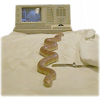
Radiography is an important diagnostic aid in reptiles. Assessment includes organ position, shape, size and density, skeletal density and gastrointestinal contents. Snakes are often presented with subcutaneous lumps and radiography is very useful to distinguish eggs from abscesses. Radiographic principles are the same as for other animals. A thorough understanding of the anatomy of the species being radiographed is essential.
Lizards
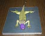
- Lizards can be radiographed with or without chemical restraint. The oculovagal reflex may be used in iguanids and other lizards may be taped down or radiographed through a box or bag if necessary.
- Positioning is very important: standard projection for dorsoventral and horiLizards can be radiographed with or without chemical restraint. The oculovagal reflex may be used in iguanids and other lizards may be taped down or radiographed through a box or bag if necessary.
Snakes
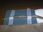
- Positioning is extremely important and snakes should not be radiographed just coiled in a bag. Radiographs of an entire snake can be taken sequentially and are far more useful than a single radiograph of coiled snake. Sequential radiographs can often be performed on a single plate. A small area of overlap should be included on each film. If possible, the lateral views should be included in the same orientation and position as the dorsoventral views.
- Barium series can be performed on snakes. Radiograph in 15 minutes to assess oesophagus, stomach and duodenum. Continue series for lower gut.
- Take two views: dorsoventral and lateral. The lateral view is the best for visualising the lung fields and the cardiac silhouette. On the DV view the spine tends to obscure the central portion of the lungs, and almost totally obliterates a clear view of the heart.
Ultrasonography
Ultrasonography is a valuable diagnostic tool and principles for mammals can be applied to reptiles. The main advantage of ultrasonography is in the evaluation of soft tissue organs. The use of ultrasonography is dependent on familiarity with the equipment, knowledge of the normal anatomy of particular species and ability to interpret the images. Minimal information has been published on the use of ultrasound for the diagnosis of diseases, but diagnostic samples and scanning for neoplasia, abscesses and cysts can be routinely performed. Ecdysis interferes with ultrasonography.
- Technique - Appropriate manual restraint is satisfactory for examining many lizards and snakes but chemical restraint may be necessary. 7.5 and 10 MHz transducers with stand-off for suitable resolution are used in small reptiles and 5 and 3.5MHz transducers for larger reptiles. The transducer (plus liberal amounts of aqueous gel) is positioned against the ventral scales. Intercostal placement is possible in large species. Initially the heart is located and the chambers and valves can be visualised. Moving caudally the liver is found as a hyperechoic organ. The stomach then spleen, pancreas and gall bladder are located caudal to the liver. Cranial to the cloaca, gonads and kidneys can be found as hyperechoic structures.
- The fat body of boid snakes can complicate the imaging of some organs including the kidneys and reproductive tract.
Echocardiography
Echocardiography is a very useful diagnostic aid for cardiac problems in species where there are well-described normals. It is non-invasive, identifies specific cardiac anatomy and quantifies cardiac function. Echocardiography can be used to evaluate heart valve motion and identify thrombi, pericardial effusion and structural defects such as stenosis and valvular disease. Presently normals for reptile species at specific temperatures are not well-described.
Endoscopy
Indications for endoscopy include:
- Diagnosis of disease by visualisation or collection of specimens,
- Treatment of disease such as the removal of foreign bodies,
- Sexing of monomorphic species,
- Assistance with other procedures such as the implanting of telemetry equipment.
Equipment
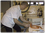
Both rigid and flexible endoscopes can be used in reptiles. A rigid endoscope is used for endoscopic coeliotomy. Expertise can be acquired by attempting endoscopy on dead reptiles and carrying out non-invasive techniques on live animals. Care is necessary since endoscopes may become contaminated with venom or Gram-negative bacteria.
Anatomy
Specific knowledge of the normal anatomy of individual species is important. The absence of an epiglottis facilitates tracheoscopy but the glottis is capable of spasm and can be easily damaged. The air sac and extensive rib cage of snakes means that insufflation is not always necessary. The anatomy of the thin, sac-like, lung means that it can be easily punctured. The long, relatively straight gastrointestinal tract facilitates examination. The vent may permit access to the colon and urinary and reproductive openings.
| Lizard and Snake Imaging Learning Resources | |
|---|---|
 Search for recent publications via CAB Abstract (CABI log in required) |
Lizard and Snake Imaging publications |
 Full text articles available from CAB Abstract (CABI log in required) |
Snake radiology: the essentials! Hernandez-Divers, S. J.; The North American Veterinary Conference, Gainesville, USA, Small animal and exotics. Proceedings of the North American Veterinary Conference, Volume 22, Orlando, Florida, USA, 2008, 2008, pp 1772-1774, 3 ref. |
| This article has been peer reviewed but is awaiting expert review. If you would like to help with this, please see more information about expert reviewing. |
Error in widget FBRecommend: unable to write file /var/www/wikivet.net/extensions/Widgets/compiled_templates/wrt699fc62af1e085_71589905 Error in widget google+: unable to write file /var/www/wikivet.net/extensions/Widgets/compiled_templates/wrt699fc62b084ff3_46185683 Error in widget TwitterTweet: unable to write file /var/www/wikivet.net/extensions/Widgets/compiled_templates/wrt699fc62b11e505_83464595
|
| WikiVet® Introduction - Help WikiVet - Report a Problem |


