Difference between revisions of "Sublingual Gland - Anatomy & Physiology"
(→Links) |
|||
| Line 16: | Line 16: | ||
<br /> | <br /> | ||
<br /> | <br /> | ||
| − | = | + | {{Learning |
| − | + | |flashcards= [[Oral_Cavity_- Anatomy & Physiology_-_Flashcards#Salivary_Glands_Flashcards|Salivary glands]] | |
| − | + | |powerpoints= [[Oral Cavity Histology resource|Histology of the Oral cavity, see part 2 for salivary glands]] | |
| + | }} | ||
Revision as of 17:11, 10 June 2011
Overview
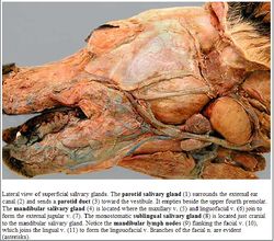
The sublingual salivary gland is a mixed gland producing both serous and mucous secretions. It is smaller than a parotid gland and consists of a monostomatic compact part drained by a single duct. It is located over the rostral mandibluar gland in the dog. The duct runs with the mandibluar gland duct and opens at the sublingual caruncle. The sublingual gland consists of a polystomatic diffuse part drained by numerous smaller ducts. There is a thin strip below the mucosa of the floor of the oral cavity and the duct opens beside the frenulum.
Histology
The gland is a tubulo-acinar gland containing mucous cells that stain lighter. The serous gland demilunes stain darker. The demilunes secrete into the lumen by canaliculi between the mucous cells.
Species Differences
Ruminant and Pig Ruminants and pigs have more mucous than serous secretions.
Equine
The diffuse part is the only part of the sublingual salivary gland present.
| Sublingual Gland - Anatomy & Physiology Learning Resources | |
|---|---|
 Test your knowledge using flashcard type questions |
Salivary glands |
 Selection of relevant PowerPoint tutorials |
Histology of the Oral cavity, see part 2 for salivary glands |
