Difference between revisions of "Hard Palate"
Jump to navigation
Jump to search
| Line 21: | Line 21: | ||
==Histology== | ==Histology== | ||
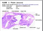
| + | [[Image:Hard Palate Histology.jpg|thumb|right|150px|Hard Palate (Mouse) - Copywright RVC 2008]] | ||
*Thick mucosa | *Thick mucosa | ||
| − | *keratinised stratified squamous epithelium | + | *keratinised stratified squamous epithelium |
| − | |||
==Species Differences== | ==Species Differences== | ||
Revision as of 13:43, 3 July 2008
Introduction
The hard palate (palatum durum) forms the rostral roof of the oral cavity. It merges caudally with the soft palate where a connective tissue aponeurosis replaces the bone.
Functional Anatomy
- Bony shelf of palatine processes of the incisive, maxillary and palatine bones. Failure of the palatine bones to fuse results in cleft palate.
- 6-8 fixed transverse ridges to direct food caudally
- Flat
- Incisive papilla (small median swelling) behind incisive teeth
- Smaller papillae ducts branching to nasal cavity and veromeronasal organ
Histology
- Thick mucosa
- keratinised stratified squamous epithelium
Species Differences
- More heavily keratinised in herbivores
