Difference between revisions of "Dermatophytosis"
Jump to navigation
Jump to search
m |
m |
||
| Line 9: | Line 9: | ||
<br> | <br> | ||
| − | + | ||
*Dermatophytes in [[Mycotic skin infections - Pathology#Dermatophytoses|dermatophytosis]] | *Dermatophytes in [[Mycotic skin infections - Pathology#Dermatophytoses|dermatophytosis]] | ||
| Line 16: | Line 16: | ||
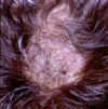
[[Image: Ringworm dog.jpg|100px|thumb|right|<small><center>Ringworm in a dog (Courtesy of Bristol BioMed Image Archive)</center></small>]] | [[Image: Ringworm dog.jpg|100px|thumb|right|<small><center>Ringworm in a dog (Courtesy of Bristol BioMed Image Archive)</center></small>]] | ||
[[Image: Trichophyton mentagrophytes dog.jpg|100px|thumb|right|<small><center>Trichophyton mentagrophytes in a dog (Courtesy of Bristol BioMed Image Archive)</center></small>]] | [[Image: Trichophyton mentagrophytes dog.jpg|100px|thumb|right|<small><center>Trichophyton mentagrophytes in a dog (Courtesy of Bristol BioMed Image Archive)</center></small>]] | ||
| + | |||
| + | Dermatiaceous fungi are pigmented, saprophytic organisms - Phaeohyphomycetes | ||
| + | *They usually infect animals secondary to traumatic implantation of the organisms, and are therefore most often seen in subcuticular or cutaneous sites. | ||
| + | *In immunuocompromised hosts they may develop systemic infections. | ||
| + | |||
| + | Phaeohyphomycosis: | ||
| + | *It occurs sporadically in cats, horses, cattle, fish, reptiles, amphibians, and birds, and rarely in dogs. | ||
| + | Fungi implicated in animal phaeohyphomycosis include: Exophiala sp., Phialophora sp., Pseudomicrodochium sp., Bipolaris sp., Moniella sp., Cladosporium sp., Wangiella sp., Curvularia spp., Exserohilum sp., Alternaria sp., Staphylotrichum sp., and Xylohypha sp. | ||
| + | *Culture is necessary for definitive diagnosis. | ||
| + | |||
*Caused by [[Fungi|dermatophytes]] | *Caused by [[Fungi|dermatophytes]] | ||
Revision as of 18:44, 28 April 2009
| This article is still under construction. |
|
|
- Dermatophytes in dermatophytosis
Dermatiaceous fungi are pigmented, saprophytic organisms - Phaeohyphomycetes
- They usually infect animals secondary to traumatic implantation of the organisms, and are therefore most often seen in subcuticular or cutaneous sites.
- In immunuocompromised hosts they may develop systemic infections.
Phaeohyphomycosis:
- It occurs sporadically in cats, horses, cattle, fish, reptiles, amphibians, and birds, and rarely in dogs.
Fungi implicated in animal phaeohyphomycosis include: Exophiala sp., Phialophora sp., Pseudomicrodochium sp., Bipolaris sp., Moniella sp., Cladosporium sp., Wangiella sp., Curvularia spp., Exserohilum sp., Alternaria sp., Staphylotrichum sp., and Xylohypha sp.
- Culture is necessary for definitive diagnosis.
- Caused by dermatophytes
- Microsporum - zoophilic
- Trichophyton - geophilic
- Epidermophyton - anthropophilic
- Common in many species, especially cats
- Hot, humid environment predisposes and viable fungi peripherally
- More common in young animals
- Produce proteolytic enzymes to penetrate surface lipid
- Fungal hyphae invade keratin -> break into arthrospores
- Epidermal hyperplasia (hyperkeratosis, parakeratosis, acanthosis) and inflammation
- Superficial perivascular dermatitis -> exocytosis (migration through epidermal layers) -> intracorneal microabscesses
- Exocytosis -> folliculitis -> furunculosis
- Highly variable lesions
- Normal -> eruptive nodular -> pseudomycetoma -> onychomycosis
- Grossly:
- Circular or irregular lesion, may coalesce
- Scaly to crusty patches
- Alopecia due to broken hair shafts and hairs lost from inflammed follicles
- Follicular papules and pustules
- Peripheral red ring (ringworm) due to dead fungi in areas of inflammation at centre of lesions and viable fungi peripherally
- Microscopically:
- Perifolliculitis, folliculitis or furunculosis
- Epidermal hyperplasia
- Intracorneal microabscesses
- Septate hyphae or spores may be found in stratum corneum and keratin of hair follicles


