Difference between revisions of "Rumen - Anatomy & Physiology"
Jump to navigation
Jump to search
m (Text replace - '|maplink = Alimentary (Concept Map)- Anatomy & Physiology' to '|maplink = ') |
|||
| Line 3: | Line 3: | ||
|linkpage =Alimentary - Anatomy & Physiology | |linkpage =Alimentary - Anatomy & Physiology | ||
|linktext =Alimentary System | |linktext =Alimentary System | ||
| − | |maplink = | + | |maplink = |
|pagetype =Anatomy | |pagetype =Anatomy | ||
|subtext1=STOMACH AND ABOMASUM | |subtext1=STOMACH AND ABOMASUM | ||
Revision as of 00:26, 3 July 2010
|
|
Introduction
The rumen is the first chamber of the ruminant stomach. It is the largest chamber and has regular contractions to move food around for digestion, eliminate gases through eructation and send food particles back to the mouth for remastication.
The rumen breaks down food particles through mechanical digestion and fermentation with the help of symbiotic microbes. Volatile fatty acids are the main product of ruminant digestion.
Structure
- Grooves correspond with thickened smooth muscle pillars on the inside of the rumen
- Ruminal pillars divide the dorsal and ventral ruminal sacs
- Coronary pillars divide the caudal blind sacs
- Cranial pillar divides the dorsal and cranial sacs
- Covered by greater omentum
- 38-40°C
- Anaerobic
- pH 6.7
- Buffered
- Large holding capacity
- Water intake lowers the ruminal temperature so bacteria are tolerant to temperature changes towards the lower end of the scale
- Objects are often lodged in the rumino-reticular fold. When the rumen contracts, the object can be pushed through the reticulum wall into the pericardium and heart.
- Laterally compressed
- Extends from the cardia at the level of the 8th rib to the pelvic inlet
- Serosa covers the entire rumen except dorsally where the rumen attaches to the abdominal roof allowing more freedom for ruminal movement and expansion
- Ruminal contractions can be felt for in the left paralumbar fossa
- 1-2 contractions should be felt per minute
- Opening at the cardia into both the rumen and reticulum is called the reticuluar groove (see oesophageal groove)
Function
- Waste removal
- Simpler products of digestion are assilimated directly, others continue down the digestive tract for further digestion
- Mixes food
- Moves food forwards through the stomach chambers
- Sensors in the rumen can determine the courseness of the food. Course, tough feed needs stronger and more frequent ruminal contractions.
- Vagus nerve (CN X) is needed for control of stomach movements
- Reflex control through sensory receptors in the medulla
- See rumination
- See eructation
Ruminoreticular contraction
- Primary mixes food
- Secondary lets gas out
- See eructation
- Ingesta flows from ventral blind sac to dorsal blind sac to dorsal sac (eructation) to ventral sac
Vasculature
- Cranial mesenteric artery
- Celiac artery
- Right and left ruminal arteries
Innervation
- Dorsal vagus (CN X) (most important)
- Ventral vagus (CN X)
Lymphatics
- Caudal mediastinal lymph node enlargement put pressure on the dorsal vagus effecting ruminal contractions
- Numerous small lymph nodes are scattered in the ruminal grooves
- The lymph drains to larger atrial nodes between the cardia and omasum, then to the cistera chyli
Histology
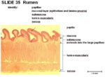
- Keratinised stratified squamous epithelium
- Non-glandular
- No lamina muscularis
- 2 thick layers of tunica muscularis- inner circular and outer longitudinal
- Interior surface of the rumen forms numerous papillae
- Papillae can be long and foliate
- Papillae can be short and pointed
- Up to 6mm in length
- Animals fed on rough grass or in the dry season have longer papillae
- Animals fed on digestible feed or in the wet season have shorter papillae (1-2mm in length)
- Fewer dorsally
- Increase surface area for volatile fatty acid absorption
- The upper keratinised layer of papillae protects against abrasion
- The deeper layers of papillae metabolise the volatile fatty acids
Rumen Microbes
- Have a variety of microbes that can utilise many substrates
- Dominance of different bacterial species depends on pH. Ergo, microbial populations are not constant
- Microbes digest cellulose and hemi-cellulose
- Microbes provide a source of all amino acids
- Microbes synthesise vitamins (especially the B vitamins)
Rumen Microbial Population
- Bacteria
- Over 2000 species
- 99.5% obligate anaerobes
- Protozoa
- Large
- Unicellular organisms
- Prey on bacteria
- Numbers affected by diet
- Fungi
- Digest fibre
- Numbers present usually low
Common Rumen Microbes
| Species | Type | pH |
|---|---|---|
| Ruminococcus flavefauens | Fibre | 6.15 |
| Fibrobacter succinogens | Fibre | 6 |
| Megashpaera eisdeni | Lactate user | 4.9 |
| Streptococcus bovis | Lactate producer | 4.55 |
Species Differences
Small Ruminats
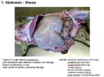
- Sheep and goats have a larger ventral ruminal sac than dorsal ruminal sac
- Cranial mesenteric artery and celiac artery come off the same root
Bovine
- Cranial mesenteric artery and celiac artery are close in the cow
- Dairy cows have a rumen pH of 5.5 due to more digestible feed
Links
The Reticulum - Anatomy & Physiology
The Omasum - Anatomy & Physiology
The Abomasum- Anatomy & Physiology
Video
Pot 52 Lateral view of the Abdomen of a young Ruminant
Pot 175 Sections of the Ruminant Stomach
Left sided topography of the Ovine Abdomen and Thorax


