Difference between revisions of "Jejunum - Anatomy & Physiology"
(→Links) |
(→Links) |
||
| Line 36: | Line 36: | ||
|flashcards = [[Jejunum - Anatomy & Physiology - Flashcards|Jejunum anatomy]] | |flashcards = [[Jejunum - Anatomy & Physiology - Flashcards|Jejunum anatomy]] | ||
|videos = [http://stream2.rvc.ac.uk/Anatomy/bovine/Pot0048.mp4 The Small and Large intestine of the Ruminant]<br>[http://stream2.rvc.ac.uk/Anatomy/bovine/Pot0052.mp4 Lateral view of the Abdomen of a young Ruminant]<br>[http://stream2.rvc.ac.uk/Anatomy/canine/Pot0036.mp4 The Canine Abdomen]<br>[http://stream2.rvc.ac.uk/Anatomy/equine/Pony_abdomen.mp4 Lateral View of the Equine Abdomen]<br>[http://stream2.rvc.ac.uk/Frean/Pony/right_topography.mp4 Right sided topography of the Equine Abdomen]<br>[http://stream2.rvc.ac.uk/Frean/sheep/LargeSmallIntestine.mp4 Small and Large intestine of the Sheep]<br>[http://stream2.rvc.ac.uk/Anatomy/swine/Pig_abdomen.mp4 The Porcine Abdomen] | |videos = [http://stream2.rvc.ac.uk/Anatomy/bovine/Pot0048.mp4 The Small and Large intestine of the Ruminant]<br>[http://stream2.rvc.ac.uk/Anatomy/bovine/Pot0052.mp4 Lateral view of the Abdomen of a young Ruminant]<br>[http://stream2.rvc.ac.uk/Anatomy/canine/Pot0036.mp4 The Canine Abdomen]<br>[http://stream2.rvc.ac.uk/Anatomy/equine/Pony_abdomen.mp4 Lateral View of the Equine Abdomen]<br>[http://stream2.rvc.ac.uk/Frean/Pony/right_topography.mp4 Right sided topography of the Equine Abdomen]<br>[http://stream2.rvc.ac.uk/Frean/sheep/LargeSmallIntestine.mp4 Small and Large intestine of the Sheep]<br>[http://stream2.rvc.ac.uk/Anatomy/swine/Pig_abdomen.mp4 The Porcine Abdomen] | ||
| + | |powerpoints = [[Gastrointestinal Tract Histology resource|Histology of the jejunum - see part 1]] | ||
}} | }} | ||
[[Category:Small Intestine - Anatomy & Physiology]] | [[Category:Small Intestine - Anatomy & Physiology]] | ||
[[Category:A&P Done]] | [[Category:A&P Done]] | ||
Revision as of 15:06, 3 June 2011
Introduction
The jejunum continues from the duodenum and leads into the ileum. It is the longest part of the small intestine and is highly coiled. It has digestive and absorptive functions.
Structure
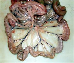
Jejunum occupies the ventral part of the abdominal cavity, filling those parts that are not occupied by other viscera. This produces species variation (see species differences). It lies on the abdominal floor, separated from the parietal peritoneum by the greater omentum. It is suspended by the mesentery (mesojejunum). This conveys the blood vessels and nerves and houses lymph nodes. The mesentery converges to its root. This is where the cranial mesenteric artery branches off from the aorta.
Vasculature
The cranial mesenteric artery, a branch of the abdominal aorta, supplies blood to the jejunum, ileum, caecum, ascending colon and part of the transverse colon. It branches greatly within the mesentery of the jejunum. There are many anastomoses within the mesentery, which ensure that the intestine can survive even if a major division of the cranial mesenteric artery is damaged. The cranial mesenteric vein drains blood from the jejunum and enters the portal vein. It is rich in the products of digestion following a meal. The portal vein enters the liver.
Species Differences
The position of the jejunum is variable between species as it lies in that part of the abdomen not occupied by other viscera.
Canine
The jejunum lies roughly symmetrically about the midline. It contacts the liver, stomach and spleen cranially and urinary bladder ventrally.
Equine
The jejunum is confined to the left dorsal part of the abdomen. It is restricted to this position by the large caecum on the right, and ascending colon ventrally on both sides.
Ruminant
The jejunum is pushed entirely to the right side of the abdomen by the rumen which is on the left. Coils of the jejunum usually lie within the supraomental recess; although this can vary between individuals depending on fullness of the rumen and size of the uterus.
Porcine
The jejunum lies in the caudoventral aspect of the abdominal cavity, mainly to the right of the midline. This is due to the presence of the ascending colon on the left.
Links
Click here for information on pathology of the Small and Large Intestines
| Jejunum - Anatomy & Physiology Learning Resources | |
|---|---|
 Test your knowledge using flashcard type questions |
Jejunum anatomy |
 Selection of relevant videos |
The Small and Large intestine of the Ruminant Lateral view of the Abdomen of a young Ruminant The Canine Abdomen Lateral View of the Equine Abdomen Right sided topography of the Equine Abdomen Small and Large intestine of the Sheep The Porcine Abdomen |
 Selection of relevant PowerPoint tutorials |
Histology of the jejunum - see part 1 |
