Difference between revisions of "Veterinary Dentistry Q&A 12"
| Line 18: | Line 18: | ||
The earliest radiologic evidence of bone loss is loss of the lamina dura (the radiologically apparent radiopaque line surrounding the tooth roots; bone loss continues in the interdental and interradicular regions with severe cortical bone loss eventually occurring. | The earliest radiologic evidence of bone loss is loss of the lamina dura (the radiologically apparent radiopaque line surrounding the tooth roots; bone loss continues in the interdental and interradicular regions with severe cortical bone loss eventually occurring. | ||
| − | |l1= | + | |l1=Hyperparathyroidism |
|q2=What is the mechanism for the development of this problem? | |q2=What is the mechanism for the development of this problem? | ||
|a2= | |a2= | ||
| Line 26: | Line 26: | ||
An alternative theory has been proposed, namely the ‘vitamin D trade-off hypothesis.’ According to this theory there is a decrease in vitamin D synthesis in the proximal renal tubules; decreased serum vitamin D levels result in loss of negative feedback on the parathyroid gland; decreased serum vitamin D levels also result in a decreased serum calcium level; an increase in serum parathyroid hormone (PTH) level occurs as a result of decreased serum vitamin D and calcium. | An alternative theory has been proposed, namely the ‘vitamin D trade-off hypothesis.’ According to this theory there is a decrease in vitamin D synthesis in the proximal renal tubules; decreased serum vitamin D levels result in loss of negative feedback on the parathyroid gland; decreased serum vitamin D levels also result in a decreased serum calcium level; an increase in serum parathyroid hormone (PTH) level occurs as a result of decreased serum vitamin D and calcium. | ||
| − | |l2= | + | |l2=Hyperparathyroidism |
</FlashCard> | </FlashCard> | ||
Latest revision as of 22:40, 13 October 2011
| This question was provided by Manson Publishing as part of the OVAL Project. See more Veterinary Dentistry Q&A. |
| Question | Answer | Article | |
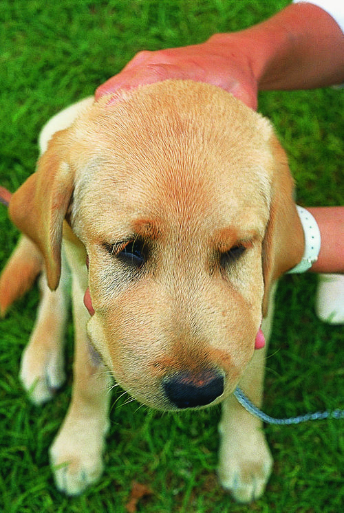
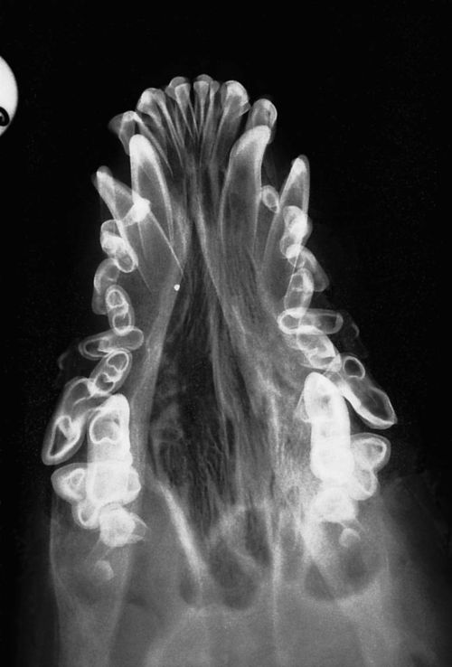
| What is the primary differential diagnosis in this eight-month-old dog on a balanced diet? | Renal secondary hyperparathyroidism. Radiologic changes in renal secondary hyperparathyroidism include generalized bone loss with a greater extent of loss occurring in the bones of the skull. The bones of the head are affected before involvement of the axial skeleton or long bones. The mandible is affected first followed by the bones in the maxilla. The earliest radiologic evidence of bone loss is loss of the lamina dura (the radiologically apparent radiopaque line surrounding the tooth roots; bone loss continues in the interdental and interradicular regions with severe cortical bone loss eventually occurring. |
Link to Article | |
| What is the mechanism for the development of this problem? | The skeletal changes occur secondary to chronic renal insufficiency, which results in secondary hyperparathyroidism. The ‘classic explanation’ is that phosphorus retention occurs as the kidneys progressively lose their ability to excrete phosphorus; phosphorus retention decreases extracellular calcium due to the mass law equation; the parathyroids are chronically stimulated to maintain extracellular calcium concentration within the normal range. An alternative theory has been proposed, namely the ‘vitamin D trade-off hypothesis.’ According to this theory there is a decrease in vitamin D synthesis in the proximal renal tubules; decreased serum vitamin D levels result in loss of negative feedback on the parathyroid gland; decreased serum vitamin D levels also result in a decreased serum calcium level; an increase in serum parathyroid hormone (PTH) level occurs as a result of decreased serum vitamin D and calcium. |
Link to Article | |

