Difference between revisions of "Avian Liver - Anatomy & Physiology"
Jump to navigation
Jump to search
| Line 7: | Line 7: | ||
*2 lobes | *2 lobes | ||
| − | *Dark brown coloured | + | *Dark brown coloured (except just after hatching where it is yellow) |
*Right lobe larger than left lobe | *Right lobe larger than left lobe | ||
| − | *Ventral and caudal to the heart | + | *Ventral and caudal to the heart (as there is no diaphragm) |
*CLosely associated to the proventriculus and spleen | *CLosely associated to the proventriculus and spleen | ||
| Line 18: | Line 18: | ||
*Indistinct lobation | *Indistinct lobation | ||
| + | |||
| + | *2 bile ducts enter the distal [[Duodenum - Anatomy & Physiology|duodenum]], one from each lobe of the liver | ||
| + | |||
| + | *The duct from the right lobe is connected to the gallbladder | ||
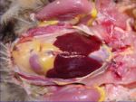
[[Image:Anatomy of the Avian Liver.jpg|thumb|right|150px|Anatomy of the Liver(Avian)- Copyright RVC 2008]] | [[Image:Anatomy of the Avian Liver.jpg|thumb|right|150px|Anatomy of the Liver(Avian)- Copyright RVC 2008]] | ||
==Function== | ==Function== | ||
Revision as of 08:33, 15 July 2008
Structure
- 2 lobes
- Dark brown coloured (except just after hatching where it is yellow)
- Right lobe larger than left lobe
- Ventral and caudal to the heart (as there is no diaphragm)
- CLosely associated to the proventriculus and spleen
- Thin capsule
- Indistinct lobation
- 2 bile ducts enter the distal duodenum, one from each lobe of the liver
- The duct from the right lobe is connected to the gallbladder
Function
- See liver funtion
Vasculature
Innervation
Lymphatics
- See liver lymphatics
Histology
- Polyhedral and angular cells
- Larger cells than in mammals
- Large, spherical nucleus
- Base of cell forms a wall of the sinusoid
- Cell apices communicate with the bile canaliculi
- Granular cytoplasm
- Liver cords form columns around the interlobular bile capillary. The cell arrangement is simpler than in mammals.
- Sinusoids anastamose freely
- Kupfer cells present
- Reticular fibres support the liver cords
- Elastic fibres in the capsule and vessels
