Difference between revisions of "Odontoblasts - Anatomy & Physiology"
Jump to navigation
Jump to search
m (Text replace - '|maplink = Alimentary (Concept Map)- Anatomy & Physiology' to '|maplink = ') |
|||
| Line 4: | Line 4: | ||
|linktext =Alimentary System | |linktext =Alimentary System | ||
|maplink = | |maplink = | ||
| − | |||
|sublink1=Oral Cavity - Teeth & Gingiva - Anatomy & Physiology#Anatomy of the Enamel Organ | |sublink1=Oral Cavity - Teeth & Gingiva - Anatomy & Physiology#Anatomy of the Enamel Organ | ||
|subtext1=ANATOMY OF THE ENAMEL ORGAN | |subtext1=ANATOMY OF THE ENAMEL ORGAN | ||
| Line 32: | Line 31: | ||
*Rate of [[Dentine - Anatomy & Physiology|dentine]] synthesis is increased during repair as it is innervated (but still acellular). | *Rate of [[Dentine - Anatomy & Physiology|dentine]] synthesis is increased during repair as it is innervated (but still acellular). | ||
| + | |||
| + | |||
| + | [[Category:Alimentary System]] | ||
Revision as of 16:44, 30 August 2010
|
|
Introduction
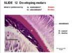
Odontoblasts are cells in the enamel organ which forms the tooth. They secrete dentine.
Properties
- Derived from mesenchyme
- Single layer of elongated columnar cells
- At dental-pulp border
- Outer layer of the dental papilla
- The first layer of dentine is formed on the enamel organ. As production increases, the odontoblasts are displaced from the enamel
- It is a major part of the tooth structure and is produced continually by the odontoblasts.
- Rate of dentine synthesis is increased during repair as it is innervated (but still acellular).
