Difference between revisions of "Lingual Gland - Anatomy & Physiology"
Jump to navigation
Jump to search
| Line 1: | Line 1: | ||
| − | |||
| − | |||
| − | |||
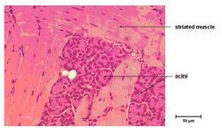
| − | [[Image:Mucous Lingual Gland.jpg|thumb|right| | + | [[Image:Serous Lingual Gland.jpg|thumb|right|250px|Serous Lingual Gland Histology (Mouse) - Copyright RVC 2008]] |
| − | + | ||
| + | Acini with cuboidal epithelium, round basal nuclei and cytoplasm that stains pink is a [[Serous Salivary Gland - Anatomy & Physiology|serous]] secreting lingual gland in the body of the [[Tongue - Anatomy & Physiology|tongue]]. | ||
| + | |||
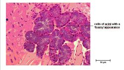
| + | [[Image:Mucous Lingual Gland.jpg|thumb|right|250px|Mucous Lingual Gland Histology (Mouse) - Copyright RVC 2008]] | ||
| + | |||
| + | Acini with flattened basal nuclei, cytoplasm that stains blue and has a foamy appearence is a [[Mucous Salivary Gland - Anatomy & Physiology|mucous]] secreting lingual gland in the root of the [[Tongue - Anatomy & Physiology|tongue]]. | ||
| + | |||
| − | |||
[[Category:Salivary Glands - Anatomy & Physiology]] | [[Category:Salivary Glands - Anatomy & Physiology]] | ||
[[Category:Oral Cavity and Oesophagus - Histology]] | [[Category:Oral Cavity and Oesophagus - Histology]] | ||
| − | [[Category:To Do - | + | [[Category:To Do - AimeeHicks]] |

