Clinical Case 9 - Page 3
Jump to navigation
Jump to search
BACK
Courtesy of C. Antonczyk
Passage of a stomach tube was attempted, but could not be accomplished.
Does successful passage of a stomach tube rule out a GDV?
- No.
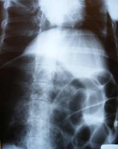
A radiograph was taken and confirms the suspected diagnosis. This radiograph was taken conscious and as the animal would not lie on its side, a dorsoventral image was taken. Note that this dog was so big the entire abdomen did not entirely fit onto the largest plate which was used here.
The caudal part of the thorax and the apex of the heart can be seen towards the top of the image. Part of the greatly distended stomach can be seen in the lower left quadrant of the radiograph.
- Click here to see a lateral radiograph of a dog with GDV.
What would you do next?
- Click here to see what the vet did.
