Equine Upper Respiratory Tract - Horse Anatomy
Nasal Cavity
Paranasal Sinuses
Overview
The paranasal sinuses of the horse are extensive, consisting of six pairs:
- Frontal and dorsal conchal sinuses (known as the conchofrontal sinus)
- Ventral conchal sinus
- Sphenopalatine sinus
- Rostral and caudal maxillary sinuses
The most clinically significant sinuses are the frontal and maxillary. The sinuses all communicate with the nasal cavity to allow drainage. The rostral and caudal maxillary sinuses communicate directly with the nasal cavity. The dorsal, middle & ventral conchal, frontal and sphenopalatine sinuses drain indirectly via the maxillary sinuses. The conchal sinuses lie within the fine, scroll-shaped bones known as conchae or turbinates. These conchae are attached to the lateral wall of the nasal passages. The paranasal sinuses are lined with respiratory epithelium (pseudostratified ciliated columnar) and goblet cells.
Frontal Sinus
The frontal sinus occupies the skull from a point midway between the infraorbital foramen and the medial canthus of the eye to a point midway between the caudal edges of the orbit. The frontal sinus is divided into right and left compartments by a midline septum. The conchofrontal sinus is formed by a communication between the rostromedial frontal sinus and the dorsal conchal sinus. The frontomaxillary aperture is a large area of communication between the frontal sinus and the caudal maxillary sinus, this is important to allow drainage. Blood supply to the frontal sinus is provided by the ethmoidal artery.
Conchal Sinuses
The main blood supply is provided by the arterial ethmoid rete, which is an anastamosis between the internal and external ethmoid arteries. A minor suply is provided by the caudal nasal branch of the sphenopalatine artery.
The conchal sinuses include the dorsal, ventral and middle. Each conchus is divided into two compartments, rostral and caudal, by a complete septum.
- Dorsal conchal sinus: This is formed by the caudal compartment of the concha
- Ventral conchal sinus: This is formed by the caudal compartment of the ventral concha
- Middle conchal sinus: Lies within the greater ethmoturbinate, not clinically significant
Maxillary Sinus
The blood supply is provided by branches of the sphenopalatine artery. This is the largest sinus and is divided into rostral and caudal compartments by a bony septum. The position of this septum is variable, but it usually lies obliquely across the roots of the 4th and 5th cheeck teeth (Tridan 109, 110, 209, 210). In horses less than 5 years of age, the reserve crown of the 3rd-6th cheek teeth (Tridan 108, 208, 109-111, 209-211) almost fills the maxillary sinus.
The rostral maxillary sinus opens via the nasomaxillary opening into the middle nasal meatus. There is also a communication between the rostral maxillary sinus and the ventral conchal sinus, via the conchomaxillary opening; located just medial to the infraorbital canal. Dorsally, there is communication with the frontal/conchofrontal sinus through the frontomaxillary opening. Between the rostral margin of the frontomaxillary opening and the conchal bulla, there is a passageway which connects the rostral and caudal compartments. This allows the caudal maxillary sinus to drain via the rostral maxillary sinus via the nasomaxillary opening into the middle nasal meatus.
Sphenopalatine Sinus
In the horse, the sphenoid and palatine sinus compartments communicate and are hence known as the sphenopalatine sinus. The sphenopalatine sinus drains via the caudal maxillary sinus, with which is communicates freely over the infraorbital canal. This sinus lies under the ethmoidal labrynth.
Guttural Pouches
Also known as: Auditory Tube Diverticulum
Introduction
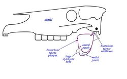
The guttural pouches are paired ventral diverticulae of the eustachian (auditory) tubes, formed by escape of mucosal lining of the tube through a relatively long ventral slit in the supporting cartilages. The auditory tube connect the nasal cavity and middle ear and the diverticulum dilates to form pouches which can have a capacity of 300-500ml in the domestic horse. The pouches are normally air filled.
Structure
The Guttural Pouch is located below the cranial cavity, towards the caudal end of the skull/wing of atlas. It is covered laterally by the Pterygoid muscles, parotid and mandibular glands. The floor lies mainly on the pharynx and beginning of the Oesophagus. The medial retropharyngeal lymph node lies between the pharynx and ventral wall of the pouches.
Right and left pouches are separated dorsomedially by rectus capitis ventralis and longus capitis muscles. Below this, by fused walls of the two pouches, the median septum is formed.
Each pouch is moulded to the stylohyoid muscle which divides the medial and lateral compartments, the medial compartment being approximately double the size of the lateral one and extends further caudally and ventrally.
The guttural pouch has close association with many major structures including several cranial nerves (glossopharyngeal, vagus, accessory, hypoglossal), the sympathetic trunk and the external and internal carotid arteries. The pouch directly covers the temporohyoid joint. The pouch has an extremely thin wall which is lined by respiratory epithelium which secretes mucus. This normally drains into the pharynx when the horse is grazing.
Several cranial nerves and arteries lie directly against the pouch as they pass to and from foramina in the caudal part of the skull (vessels within mucosal folds that indent the pouches):
Medial Compartment:
- Cranial nerves IX, X, XI, XII.
- Continuation of the sympathetic trunk beyond the cranial cervical ganglion.
- Internal carotid artery.
Lateral Compartment:
- Cranial nerve VII - limited contact with the dorsal part of the compartment.
- External carotid artery crosses the lateral wall of the lateral compartment in its approach (as maxillary artery) to the atlas canal. The external maxillary vein is also visible.
Drainage:
Natural drainage of the pouch is throught the slit-like (pharyngeal) openings of the eustachian tube in the lateral wall of the nasopharynx. The connection opens when the horse swallows and grazing normally provides drainage. However, most of the pouch is ventral to his slit, and therefore drainage may be rather ineffective. If blocked, secretions accumulate and the pouch distends producing a palpable swelling.
Function
The function of guttural pouches is largely unknown, however hypotheses have been put forward:
- It may influence internal carotid artery blood pressure. Air pressure varies with phase and forcefulness of respiration and the artery in the mucosal fold is exposed sufficiently to be affected.
- Cerebral blood cooling mechanism. Operates at times of physical stress/exercise, when core body temperature is raised. Vigorous respiration, cool air in guttural pouches, and the exposed artery all lead to cooling of the blood.
Links
Larynx
Overview
The larynx is situated below where the pharynx divides into the trachea and the oesophagus. It is contained partly within the rami of the mandible and extends caudally into the neck. It is important during breathing, vocalisation and deglutition (swallowing). The cartilagenous larynx can be manually palpated in the living animal and is commonly implicated in respiratory conditions such as roaring.
The pharynx is located rostrally to the larynx, whilst the trachea is located caudally. The larynx is suspended from the hyoid apparatus. It is bilaterally symmetrical and 'tube-shaped' and can be described as a musculocartilagenous organ.
Laryngeal Cartilages
Thyroid Cartilage
The thyroid cartilage is a hyaline cartilage and forms most of the floor of the larynx. The fusion of the two lateral plates varies in different species. The rostral part forms the 'Adam's apple'. The thyroid cartilage articulates with the thyrohyoid bone and the cricoid cartilage. It becomes brittle as the animal ages.
Arytenoid Cartilage
The arytenoid cartilages are paired, triangular shaped hyaline cartilages. They lie either side of the cricoid cartilage and articulate with the rostral part of it. A vocal process is present on the caudal surface where the vocal folds attach; a muscular process extends laterally and is the origin of attachment for the cricoarytenoideus dorsalis muscle. A corniculate process extends dorsomedially.
Epiglottic Cartilage
The epiglottic cartilage is an elastic cartilage, which is the most flexible and most rostral type of cartilage. The thinner stalk-like part, is attached to the root of the tongue, the body of the thyroid cartilage and the basihyoid bone. The larger blade-like part lies behind the soft palate and points dorso-rostrally. During deglutition, the large blade part of the epiglottic cartilage partially covers the entrance to the trachea.
Vocal Folds
The vocal folds are made of (slightly stiffer) elastic ligaments and pass between the arytenoid cartilages and the laryngeal floor. They run caudodorsally, with the ligament positioned medially and the vocalis muscle laterally. Fat surrounds the vocalis muscle. The vocal folds form part of the glottis and secrete mucous. They are used for vocalisation.
Vestibular Folds
The vestibular folds are made of (slightly stiffer) elastic ligaments. The vestibular ligaments are rostral to the vocal ligament. The vestibular folds run caudodorsally, rostral to the vocal folds with the ligament positioned medially and the vocalis muscle laterally.
Intrinsic Musculature
The intrinsic laryngeal muscles produce changes in caliber of the rima glottis by abducting and adducting the corniculate processes of the arytenoid cartilages and the vocal folds and hence, altering airway resistance. The principle muscle to widen the glottis is the cricoarytenoideus dorsalis. The thyroarytenoideus, arytenoideus transversus, and the cricoarytenoideus lateralis muscles adduct the corniculate processes of the arytenoid cartilages, narrowing the glottis and therefore protecting the lower airway during swallowing.
Cricothyroid muscle
The cricothyroid muscle is innervated by the cranial laryngeal nerve, a branch of the vagus nerve (CN X). It moves the cricoid and arytenoid cartilages caudally to tense the vocal folds.
Dorsal cricoarytenoid muscle
The dorsal cricoarytenoid muscle is innervated by the caudal laryngeal nerve, a branch of the vagus nerve (CN X). It runs from the dorsal surface of the cricoid cartilage to the arytenoid cartilage. It abducts the vocal process and therefore the vocal fold to widen the glottis and is the principle abductor of the larynx.
Lateral cricoarytenoid muscle
The lateral cricoarytenoid muscle is innervated by the caudal laryngeal nerve, a branch of the vagus nerve (CN X). It adducts the vocal processes and narrows the glottis.
Thyroarytenoid muscle
The thyroarytenoid muscle is innervated by the caudal laryngeal nerve, a branch of the vagus nerve (CN X). It runs from the laryngeal floor to the thyroid cartilage and arytenoid cartilage and alters the tension of the vocal and vestibular folds. It forms part of the sphincter muscular arrangement.
Transverse arytenoid muscle
The transverse arytenoid muscle is innervated by the caudal laryngeal nerve, a branch of the vagus nerve (CN X). It completes the muscular sphincter arrangment and spans the arytenoid cartilages.
Pharynx
Trachea
References
Dyce, K.M., Sack, W.O. and Wensing, C.J.G. (2002) Textbook of Veterinary Anatomy. 3rd ed. Philadelphia: Saunders.
