Ornamental Fish Q&A 09
| Question | Answer | Article | |
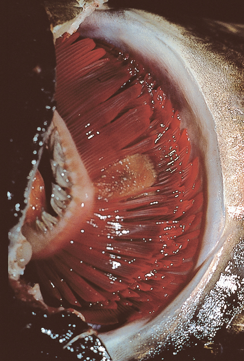
| Describe the disease condition illustrated by the photograph. | The photograph shows a discrete, oval, raised, plaque-like lesion on the gill of a koi carp recently imported to the UK from Japan. These changes were consistent with a diagnosis of branchiomycosis (gill rot), which is a disease of freshwater fish caused by the fungus Branchiomyces. It has been described in Europe, Japan, India, and parts of the USA. Spores attach to the gill surface and germinate to form hyphae. These hyphae proliferate, causing damage to the blood supply and necrosis. Sloughing of this necrotic tissue releases spores into the water, which then continue to develop on the floor of the pond or aquarium if conditions are favorable (i.e. temperatures of 25–32°C (77.0–89.6°F), high levels of organic material, low oxygen levels, and a low pH). |
Link to Article | |
| What tests can be performed to confirm the diagnosis? | The diagnosis of branchiomycosis could be confirmed by examining fresh scrapings taken from the lesion and by histopathology. In this particular case, the scraping would be expected to yield branched aseptate hyphae. Large areas of necrosis containing fungal spores and hyphae can usually be seen on histopathology with a GMS (Gomori methenamine silver) preparation. |
Link to Article | |
| How is the organism transmitted, and what measures would you advise to treat and prevent the problem? | Fungal spores can be introduced directly by adding infected or carrier fish, or indirectly by birds or the use of raw fish products. Treatment is generally unrewarding, although 2-phenoxyethanol has been suggested. Control is achieved by good hygiene, avoiding the use of raw fish products, and adequate quarantine of all new fish to avoid introduction of the organism. |
Link to Article | |
