Snake Physical Examination
| This article is still under construction. |
Introduction
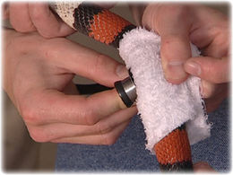
The physical examination involves observation of the snake, taking measurements and a thorough methodical area by area examination. Many techniques are similar to other animals, but before examining the snake ask the owner if it is accustomed to being handled. See here for information on handling and restraint of snakes. A veterinarian who is inexperienced with reptiles may be likely to focus on the presenting signs but then can end up treating exclusively the secondary problems. Stomatitis and rectal prolapse are secondary conditions where a full examination with husbandry review, including housing and nutrition, is vital in determining the principal problem.
Observation
Snakes should have active tongues that are sampling scent particles in the atmosphere. Normal movement should be observed although allowances must be made for any chilling effect in transit since this will reduce the patient's metabolism and give a misleading impression of lethargy and lack of strength. The righting reflex should be tested since poor reactions can be a result of weakness and not necessarily neurological disease. Respiration, including rib movements and any sounds produced. Be aware of hissing noises arising from agitation that are not a result of respiratory infection.
Body condition
Weight of the snake should be taken at every examination, as should snout-vent lengths. Obese snakes have increased fat deposition on the last third of the body before the cloaca. Cachectic snakes with significant loss of muscle mass show a prominant dorsal spine and ribs, and the body shape looks more triangular in cross section.
Skin
Examine under a bright light and with a hand lens if necessary.
Examine for:
- Ectoparasites
- Trauma due to fighting, mating or thermal burns
- Discoloration (previous scars, injection sites)
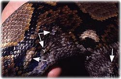
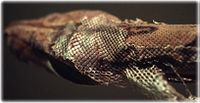
- Dysecdysis - retained skin is often dry and brown while normal ecdysis occurring in a piecemeal fashion is flexible and transparent. Retained skin is a focus for infection and acts as a tourniquet on the tail tip and digits leading to ischaemic necrosis
- Examine the scales for loss, scabs, blisters and haemorrhages, ensuring the ventral scales are inspected
- Assess for abnormal masses, swellings or bruised areas
- Assess skin elasticity
- Skin folding indicates dehydration, cachexia. Assessment of dehydration in reptiles is analogous to mammals. With 5-8% dehydration there is loss of skin elasticity with a wrinkled appearance. With more severe dehydration (10-15%) the eyes are sunken and the mucous membranes become dry and sticky (consider that the oral cavity of most reptiles is relatively dry compared with mammals).
Find out more about snake skin
Find out more about snake skin diseases
Shedding
Ecdysis (shedding) generally takes place every 14 days although this is dependent on many variables such as environment, age and size of snake. Snakes shed their entire skin in one piece, including the spectacles, although large snakes greater than 3m in length may shed their skin in incomplete sections. Prior to ecdydsis, a snake will become anorectic and handling may be hazardous to the animal at this time if the underlying epidermis is damaged.
Head
Nares
The nares should be clear and free of discharge and retained shed.
Eyes
The eyes are usually considered as a barometer of general health and environmental conditions; therefore a full ophthalmologic examination should be performed in all cases.
- Following a general assessment of the eye, a more detailed examination should be performed using a form of focal illumination and a magnification system.
- Examination of the ocular fundus to assess the retina (fundoscopy) can be carried out using a direct or indirect ophthalmoscope.
- Mydriasis can be attempted either through general anaesthesia or intracameral injection of neuromuscular blocking agents such as curare or d-tubocurarine. These agents can also be applied topically; however their effectiveness may be affected by the variable corneal penetration of the drugs.
- Other diagnostic tools, such as tonometry (measurement of the intraocular pressure), stains, cytology, bacteriology, histopathology and electron microscopy, in addition to routine diagnostic tools (haemotology, biochemistry, radiology and ultrasound) can also be used to detect an ocular disease or underlying problem in snakes.
- Retained spectacles are a concern. They should be clear with no signs of subspectacluar disease.
Oral cavity
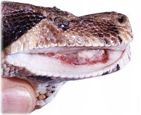
- The oral cavity should be opened using a soft, pliable speculum
- Mucous membranes should be pale to pink and free of thick ropey mucous
- Evaluate the tongue for movement
- The glottis should be gree of discharge
- Examine the teeth for fractures
References
Fowler, M.E. and Miller, R.E. (2003). Zoo and Wild Animal Medicine. Saunders, 5th Edition. pp. 84. ISBN 0-7216-9499-3


