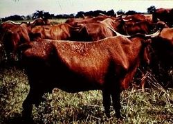
Dermatophilosis
| This article is still under construction. |
| Also known as: | Cutaneous streptothrichosis Lumpy wool - sheep Strawberry foot rot - sheep |
Description
This is a group of diseases affecting the epidermis caused by Dermatophilus congolensis. It causes a range of conditions in large animals including rain scald in horses and strawberry foot rot in sheep. The disease is associated with skin trauma, prolonged wetting or parasites. Lesions typically involve exudative dermatitis with scab formation. It is a zoonosis affecting humans in close contact with infected animals.
Signalment
Can be seen in animals of all ages but most commonly occurs in young animals who are chronically exposed to moisture. Affects horses, sheep, cattle, goats, pigs and rarely dogs and cats.
History and Clinical signs
Lesions commonly occur following heavy rainfall and commonly affects the dorsum of animals. Any previous trauma or damage to the skin can predispose to infection. Blood-sucking insects are also thought to be involved in transmission.
Bovine dermatophilosis
Also see General Dermatophilosis
Is rarely reported but causes lesions which are distributed over the head, dorsum, neck and chest. Cattle that stand for long periods in deep water and mud develop lesions over the flexor surfaces of the joints. Dairy cows may develop lesions on the udder.
Lesions may resolve within weeks if dry weather or prolonged wetting of infected areas can lead to secondary bacterial infection which can result in limb oedema and cellulitis.
Treatment
Farm animals: Bring affected animals into a dry environment. Investigate any underlying problems which may predispose to the infection. Antibiotics can be given intramuscularly and typically work following one dose. However if signs do not resolve a 5 day course should be administered. Penicillin and streptomycin are good choices for this disease. Additionally Dips containing 0.2% Copper Sulphate or 0.5% Zinc sulphate can be effective.
Diagnosis
Can often make a diagnosis on history and physical exam. Impression smears can also be useful when stained with either gram stain or Giemsa and examined microscopically. Additionally it is possible to culture material from the crusts however this can be difficult due to the slow growing nature of the pathogen.
Pathology
Grossly: Papules, pustules, crusts may coalesce and mat the coat.
Microscopically:
- Hyperplastic superficial perivascular dermatitis
- Multilaminated crusts, alternating keratin and inflammatory cell layers
Prognosis
Good if animals are kept dry. Often re-occurs in wet weather.
References
Merck & Co (2008) The Merck Veterinary Manual (Eighth Edition) Merial
4th year Veterinary Dermatology notes. Royal Veterinary college. October-November 2008. p60-64.
