Introduction
A thrombus is a localized or generalized intravascular or intracardiac blood clot. Thrombosis is the method of formation of a thrombus. Thrombi may occur in any vasculature in the body and form as the result of trauma or pathological processes affecting the blood vessel endothelium or disturbances to blood flow and/or blood composition. Some diseases such as infective endocarditis and heart worm increase the risk of thrombi formation.
It may occur due to endothelial injury, altered blood flow e.g. abnormal stasis or hypercoagulability.
Thrombosis is often associated with other disease processes such as disseminated intravascular coagulation (DIC). In cats with hypertrophic cardiomyopathy, thrombosis often causes posterior paralysis in these animals, as it lodges in the branching of the abdominal aorta.
Spontaneous venous thrombosis is rare, but can be seen in cattle with traumatic reticulo-peritonitis in the caudal vena cava.
Types
There are many different types of thrombosis.
Arterial thrombosis is common in man, but it is uncommon in domestic animals; in man it is associated with atherosclerotic vascular disease. When they do occur in animals, arterial thrombi are usually small lesions but these may be sited strategically, thereby causing problems.
Arteriosclerosis is seen in aged dogs and horses affecting the coronary artery and other major arteries.
Verminous arteritis may occur with or without aneurysm and is seen in horses as a result of Strongylus vulgaris infestation. It affects the root of cranial mesenteric artery, renal artery and aorta as a consequence of larval migration in the vessel walls.
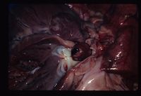
Cardiac thrombosis is usually valvular, but can occasionally be mural. In farm animals, and rarely in the horse, infective/inflammatory thrombosis occurs subsequent to endocarditis. In dogs and horses, cardiac thrombosis is generally of degenerative/non-infectious cause; endocarditis may occur, though uncommonly.
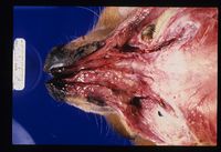
Venous thrombosis is a fairly common type of thrombus in the veterinary species because veins are relatively thin-walled and are therefore more susceptible to distortion, inflammatory damage and iatrogenic venepuncture damage. Also,veins have relatively slower blood flow rates allowing cell aggregates to persist more readily. Most venous thrombosis in domestic animals results from extension of inflammatory reactions, erosion/disruption caused by malignant tumours, pressure from adjacent space-occupying masses or venepuncture damage.
Capillary thromboses are microthrombi that are only appreciable histologically. Their formation may be localised and associated with acute local inflammation or generalised. Localised microthrombi are rarely significant unless they are strategically sited. Generalised microthrombi may be seen in terminal diseases as a reflection of vascular failure, and are often associated with shock syndromes as part of disseminated intravascular coagulation. Clinically, generalised capillary thrombosis is highly significant.
Signalment
Can occur in any specie of animal, but is most commonly described in dogs and cats.
Clinical Signs
Signs depend on the area affected and the size of the vessel blocked. There will always be poor perfusion below the affected area, or malfunction and necrosis of the affected organs. The area affected will often appear pale, cold, have poor perfusion and be painful to touch.
Diagnosis
History and clinical signs can be indicative of the condition. On blood tests, one may see abnormalities associated with lack of blood perfusion in pathological conditions that can cause the thrombosis.
Ultrasonography may show blood stasis and may also demonstrate the presence of a thrombus.
Angiography may show lack of opacity in the affected region.
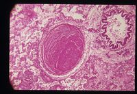
On microscopic examination, the thrombus appears as a layered mass which is attached to the vessel wall. The composition consists of red blood cells, neutrophils and platelets bound together by fibrin. Thrombi take different appearance depending on whether they are arterial or venous. Arterial thrombi tend to be pale and have a tail in the direction of blood flow. The high rate of blood flow sweeps red cells away - the thrombus is composed of mainly white cells, platelets and fibrin which are left behind. Venous thrombi tend to be a darker red. The slow blood flow allows the clot to form quicker which is loosely arranged and contains many red blood cells.
Treatment
The mainstay of treatment is to diagnose and treat the underlying problem.
It is important to give pain relief and IV Fluids as palliative care. The use of an anticoagulant such as heparin for short-term treatment or aspirin for long-term treatment should be considered.
Prognosis
Prognosis depends upon the underlying condition.
If the animal survives the immediate effects of a thrombus, the thrombus may evolve in one of the following ways:
- The thrombus may gradually enlarge and eventually cause total obstruction of a vessel.
- The thrombus may be completely removed by fibrinolytic activity; fibrinolysis is a very active process and clots are usually removed within a few days of formation provided that blood flow is sufficient. An occlusive thrombus may prevent the necessary enzymes from reaching the clot but contraction of fresh clots under the influence of thrombasthenin (released by platelets) may provide a slit-like channel beside the thrombus that permits the blood flow to restart and completely dissolve the clot.
- Organisation; a thrombus acts as a foreign body, causing an inflammatory response in the underlying blood vessel or heart wall. The external surface of the thrombus then quickly becomes covered by endothelium and is excluded from the clotting mechanism. Neutrophils invade the mass and may reduce the centre. Occasionally, subsequent invasion by bacteria may lead to purulent inflammation. Normally, fibroblasts and capillary buds follow the neutrophils into the thrombus and a fibrous vascularised connective tissue forms. Capillaries channels anastomose to produce vessels that traverse the thrombus and re-establish blood flow - this is known as canalisation of a thrombus. Fibrous tissue matures and contracts, eventually causing the thrombus to become incorporated into the vessel wall as a fibrous lump. A piece of the thrombus may break off and form an embolus.
Also see: Thromboembolosm.
References
Ettinger, S.J. and Feldman, E. C. (2000) Textbook of Veterinary Internal Medicine Diseases of the Dog and Cat Volume 2 (Fifth Edition) W.B. Saunders Company
Ettinger, S.J, Feldman, E.C. (2005) Textbook of Veterinary Internal Medicine (6th edition, volume 2) W.B. Saunders Company
Fossum, T. W. et. al. (2007) Small Animal Surgery (Third Edition) Mosby Elsevier
Merck & Co (2008) The Merck Veterinary Manual (Eighth Edition) Merial
Nelson, R.W. and Couto, C.G. (2009) Small Animal Internal Medicine (Fourth Edition) Mosby Elsevie
| This article has been peer reviewed but is awaiting expert review. If you would like to help with this, please see more information about expert reviewing. |


