Theileria
| Theileria spp | |
|---|---|
| Kingdom | Protista |
| Phylum | Protozoa |
| Order | Piroplasmorida |
| Family | Theileriidae |
| Genus | Theileria |
| Species | Theileria parva and others |
Introduction
Theileria species are a group of protozoan pathogens causing severe lymphatic proliferative disease in cattle.
T. parva is the species of most veterinary importance, affecting cattle in Central and Eastern Africa.
Other species cause significant economic losses in the Mediterranean, Middle East and Northern Africa.
Lifecycle
Theileria are transmitted via the Haemaphysalis and Rhipicephalus and Dermacentor species of tick vectors.
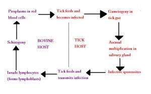
Sporozoites enter mononuclear cells of the host and develop into trophozoites and multinucleate schizonts by asexual reproduction. This process stimulates proliferation of the host cells, allowing further multiplication of the parasite. The local lymph nodes are first infected.
Schizonts then disseminate' through the ' before differentiating into merozoites.
The merozoites enter the erythrocytes and form piroplasms which are infective to ticks and capable of sexual reproduction.
Sexual reproduction occurs within the nymph and larval stages of the tick and the final infective stage is present within the salivary glands and is transmitted to mammalian hosts when bloodfeeding.
Transmission in the tick is then trans-stadial.
In endemic areas, endemic stability is often reached, in which most or all cattle may be infected and be carriers and most ticks are also infected, but young calves gain solid immunity from their immune dams and therefore rarely show clinical disease. This state however takes time to stabilise and will cause significant economic losses in the process.
For more information on ticks as vectors, see Tick Disease Transmission.
Pathogenesis
Lymphocytes are killed by invading protozoa and later in disease, lymphopoeisis is reduced and prevented.
Diseases
Theileria parva
Also Known As T. mutans and T. sergenti.
Primarily a parasite of African buffalo.
Transmitted by a wide range of tick hosts and also the burrowing mite, Sarcoptes scabei.
The cause of Bovine Theileriosis and East Coast Fever
Forms rod shaped piroplasms within host erythrocytes.
Shows extreme antigenic diversity across its geographical distribution, although parasites isolated in different diseases are genetically identical.
Can also infect sheep and mice.
Theileria annulata
Also Known As T. dispar
Also a cause of Bovine Theileriosis
Infects macrophages and B Lymphocytes.
Forms round or oval piroplasma within host erythrocytes.
Also infects sheep and yaks
References
Animal Health & Production Compendium, Theileria datasheet, accessed 04/06/2011 @ http://www.cabi.org/ahpc/
