| This question was provided by Manson Publishing as part of the OVAL Project. See more Reptiles and Amphibians Q&A. |
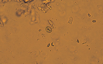
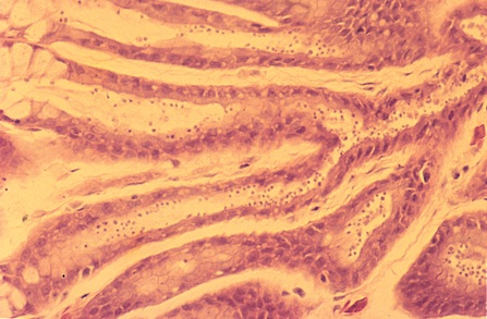
The photomicrograph is of a merthiolate-stained wet mount made from fluid recovered from the stomach of a snake after a gastric lavage. An H&E-stained histological section of the gastric fundus from the same snake is also shown.
| Question | Answer | Article | |
| What is the small organism in the middle of the microscopic field in the first picture? Myriad numbers of the same organism are seen attached to the cellular borders of the gastric pits in the second picture. | Cryptosporidium serpentis. |
Link to Article | |
| What is the prognosis? | Very guarded to poor because, to date, there appears to be no effective treatment for this severe parasitism in reptiles; the drugs most often used to treat human cryptosporidiosis have not been effective in reptiles. |
Link to Article | |
| How would you manage this condition in a colony of snakes? |
|
Link to Article | |

