Wound Management Basics - Donkey
| This article has been peer reviewed but is awaiting expert review. If you would like to help with this, please see more information about expert reviewing. |
Minimizing the potential problems of a wound

‘Time spent in the preparation of a wound is never wasted’ – Barrie Edwards, 1984
Harmful effects can be minimised by careful wound management and sound surgical techniques including:
- Early intervention
- Bacterial adhesion occurs around four to eight hours after wounding and therefore intervention before this occurs provides a much cleaner wound
- Long delays in attention to a wound inevitably result in overt infection and contamination by foreign matter
- Delay in wound examination may, however, make recognition of nonviable tissue easier
- The application of sound surgical principles
- The use of appropriate debridement techniques
- The use of suitably placed surgical drains - vacuum drains and Penrose (capillary) drains
- Minimizing dead space within the wound itself
- The use of physiologically sound wound lavage mechanisms (see Initial cleaning below and Foreign body).
- Reducing and controlling infection
- Eliminating and preventing contamination and continuing trauma
Summary
Recognition of potential problems, that is, factors that might inhibit wound healing (see Delayed primary healing), allows decisions on the best or most appropriate management and the likely results of healing. Owner and veterinary expectations can be rationalized and explained. Consideration of the problems from the outset will almost always result in earlier and more satisfactory healing. By the nature of their location and severity, many wounds will have particular limitations and needs, and these must be addressed from the outset of wound management. All wounds must be promptly and thoroughly examined to establish the full extent of the damage. It will usually be difficult to perform a full examination on a wound without appropriate sedation (or, if necessary, anaesthesia). The exact site, depth and direction of the wound are critical factors in all wound management procedures. The cause of the wound and the time delay between injury and veterinary attention will have important implications for the subsequent management. The tetanus vaccination status should always be determined and appropriate protection ensured. Sedatives, opioid analgesics with non-steroidal anti-inflammatory drugs make initial assessment and subsequent procedures far easier.
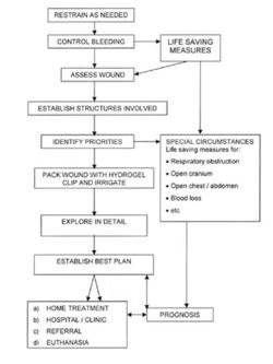
Initial procedures
1. Control of haemorrhage
This is a major initial consideration.
Arterial bleeding
Arterial bleeding is potentially the most dangerous haemorrhage. Even small arteries can produce significant blood loss. This is bright red and under high pressure.
- Control of arterial bleeding is usually effected by application of direct pressure over the site (or in the arterial tree on the heart side of the injury). This may need to be maintained for up to 10 to 15 minutes
- Alternatively a pressure bandage can be applied. It is possibly helpful to use a firm roll of bandage over the actual site to increase the local pressure without having to make the bandage dangerously tight for other tissues
- Management of pressure bandages is important. If after two hours bleeding is still present when the bandage is removed, an alternative method of haemostasis should be used
Note:
- Pressure bandages can be catastrophic without the correct bandaging technique. It may stop the bleeding but leave the donkey with extensive skin necrosis or, even worse, tendon necrosis
- Pressure bandages should not be left on for more than one to two hours
- Removal of the dressing can reinitiate bleeding
Direct ligation or clamping of the bleeding artery is of course an effective
method of control, but arteries usually run alongside nerves and inadvertent
clamping of the nerve in a conscious donkey will usually cause a dangerous
response. Also, ligation of a major end-artery can be catastrophic. Ligation
of a bleeding artery will also leave a suture in the wound and the material
used must be chosen with care.
Venous bleeding
Venous bleeding is usually dark in colour and has a 'slow flow rate, although the overall volume can be large if large veins are involved.
- Venous bleeding can be controlled by direct pressure, by application of a firm (but not tight) bandage or by allowing a normal clot to form in the site
- Usually venous bleeding will stop naturally within 10 to 15 minutes. Disruption of the clot may, however, restart the bleeding
Capillary bleeding
Capillary bleeding is slow and of low volume but can be bright or dark in colour. It can be controlled by allowing natural clotting to take place or by application of cold compresses or dressings. Capillary ooze may continue for some time after injury but is unlikely to be of sufficient volume to cause problems.
2. Initial cleaning
The first objective is to expose the injury so that a thorough assessment can be made. This will always involve clipping of the hair and removal of gross contamination. During this stage the wound itself should be covered or filled with an inert, water-soluble jelly such as obstetrical lubricant (K-Y Jelly) or hydrogel (Intrasite Gel ). This will prevent clipped hair and other superficial debris from entering the wound during clipping and initial washing of the surrounding skin.
Once the wound area is clipped and cleaned, the gel within the wound is washed out with (sterile) saline under minimal pressure (3-5 psi), but (warm) running water is commonly used until any gross contamination is dislodged. The final wash should be with normal saline to restore physiological status. Care should be taken to ensure that this does not drive foreign matter into the depths of the wound. If the wound has bled heavily, washing may loosen the blood clot and restart haemorrhage, which may then need to be controlled.
A solution of 0.5% chlorhexidine is a standard wound antiseptic with minimal harmful effects. It can be used if the wound is heavily contaminated or is over two to four hours old. Fresh wounds probably do not need an antiseptic wash.
At this stage the wound can be lavaged with warm (sterile) saline under increased pressure (7-10 psi - simply using a 50 ml syringe and squirting the saline directly from it with moderate pressure can achieve this). Higher pressure can drive bacteria and particles into the tissues and open fascial planes, while lower pressures may fail to dislodge foreign matter and bacteria.
Avoid
- Wiping the wound with dry or saline-soaked swabs that may just push bacteria and foreign matter deeper into the wound
- Strong chemical disinfectants and antiseptics. These should not be used without considerable thought on the possible balance between benefit and harm
3. Wound assessment
The wound must be fully assessed to establish its full extent and the various structures in the vicinity must be considered individually. A sterile gloved finger is a sensitive and sensible instrument to use! Sedation may be required. Small foreign bodies, bone fragments and tissue damage can be appreciated.
Major factors that need to be established in a wound
- Depth of the wound
- The direction of the damage
- The extent of the damage
- The precise tissues and structures involved
After assessment, the wound is carefully flushed again with sterile saline. The presence of any of the recognized factors that might hinder, delay or prevent healing must be recognised early. Where delayed healing is unavoidable, the owner can be advised accordingly and a suitable plan instigated to expedite the healing as much as possible without unrealistic expectations.
4. Prevention of further injury and contamination
A hydrogel or sterile water-soluble (obstetrical) lubricant gel is packed into the wound and a suitable protective dressing applied, taking care not to cause further damage.
5. Infection control

Up to six to eight hours after injury, a wound is usually considered contaminated and antibiotics could be considered to be prophylactic. Beyond six to eight hours bacteria have usually become established in the damaged tissues and the wound is then classified as infected. Antibiotics used at this stage are therapeutic. Although this so-called ‘golden period’ is an important concept, it suffers from being too prescriptive; in some cases the wound may become infected slower or faster. The overriding principle of wound management is that the wound should be dealt with as soon as possible after injury.
Antibiotics are used to treat known or suspected infections and as prophylaxis for various types of medical and surgical procedures. Notwithstanding the consideration of contamination and established infection, there is merit in administering a full dose of antibiotic before any interference is undertaken, the wound site covered throughout the procedure. Topical antibiotics are probably not helpful, but sometimes soluble antibiotic is usefully added to lavage solutions (especially in special or complicated wounds, such as wounds involving joints and body cavities).
Failure to control potential and actual infection will inevitably result in retarded healing. Removal of bacteria before adhesion occurs is a useful aid to wound healing and minimises the use of antibiotics. The side effects of antibiotics include the development of bacterial resistance, anaphylactic reactions, overgrowth of bacteria and gastrointestinal disturbances.
Important note Antibiotics seldom eliminate infection – rather they reduce the rate of bacterial replication to a degree that allows the hosts’ immune system to eliminate the infectious agent.
The donkey is particularly sensitive to tetanus toxins and the untreated disease carries a hopeless prognosis. Therefore, the tetanus vaccination status should be established in all cases. If the donkey has had a recent vaccination then there should be no risk of the disease (the vaccine is highly effective) but, where vaccination history is dubious, either a tetanus toxoid booster vaccination or antiserum (or both) should be administered.
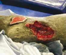
6. Wound debridement


Ideally, all foreign matter and necrotic or non-viable tissue should be removed, using a scalpel and dissecting forceps, to convert an accidental wound into a surgical one that can be closed by first intention. Although this ideal is seldom achieved it is an important target. Debridement of contaminated or devitalized tissue should be accomplished systematically, starting at the most dependent part of the wound so that bleeding does not conceal tissue that should be removed. Repeated partial debridement can be performed to produce a clean, healthy wound site. Surgical debridement may be delayed until it is possible to differentiate between viable and devitalised tissue. Extensive debridement may require general anaesthesia – this has major advantages in terms of accuracy and detail but circumstances will vary.
Important note In anatomical sites that have little ‘spare’ skin (e.g. the distal limb regions and the face) or where skin deficits are likely to have serious limiting effects (e.g. the eyelid), skin should be preserved as far as possible.
As a general rule, skin should be preserved unless its presence is likely to do more harm than good. Even non-viable skin can act as a useful (if temporary) biological cover for wound site.
Antibiotics can never replace good wound management principles. The inability to create a completely sterile wound by debridement and lavage can be partially (but NOT totally) compensated for by antibiotics applied locally and administered systemically. Provision of adequate drainage (through placement of drains), partial suturing and counter incisions to reduce fluid and tension at the wound site can also help to limit the chances of infection.
7. Wound closure
The choice of wound closure method is governed by the nature and site of the wound and is a matter of clinical preference. Wounds subjected to primary wound closure or first intention healing will usually heal faster than those subjected to either delayed primary healing or second intention healing.
Incised wounds frequently lend themselves to suturing. Suturing should only be carried out when so doing will have a positive advantage and minimal harmful effects. Careful selection of suture patterns will make a considerable difference to wound healing. No wound should be completely closed unless the deeper tissues are effectively sterile.
Factors that are likely to result in wound breakdown (dehiscence) after suturing include:
- Gross contamination, including hair matting over the wound.
- Infection.
- Significant skin loss (either occurring at the time of injury or arising afterwards from skin necrosis).
- Marked swelling with fluid and inflammatory exudate.
Proper preparation and careful assessment should limit these factors significantly.
Important note
- Delays in closure may result in contraction of the skin flaps and so preclude closure
- Primary closure will almost always fail when tissue necrosis and swelling disrupt the suture line
- Notwithstanding the presence of obvious complication factors, wounds involving the lower parts of the limbs usually present the greatest challenges. There is considerable controversy over the need/necessity to suture lower limb wounds. In general, a limb wound may be sutured if the wound is:
- Clean.
- Free of complicating factors.
- In the longitudinal plane (i.e. running up/down the limb).
- In a suitable site that makes suturing without undue tension feasible.
- Otherwise it is probably best to use second intention healing or delayed primary intention healing
Delayed primary closure may be applicable in relatively clean but contaminated wounds with extensive tissue damage. The wound is cleaned, debrided and dressed with a hydrogel and a polymeric foam dressing applied. Daily re-examination and redressing continues until the wound is free of obvious infection and necrotic tissue, and the wound bed contains healthy granulation tissue. The wound is then freshened using careful, superficial, sharp debridement and closed using a suitable suture technique (possibly with the aid of tension-relieving quills or tension-relieving lateral incisions).
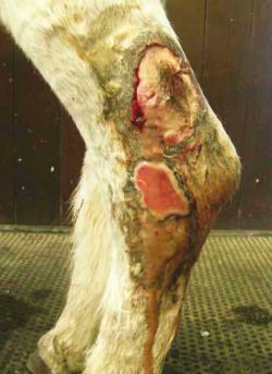
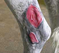
Second intention healing is applicable to the large majority of wounds in donkeys. The wound is left open after initial treatment and allowed to granulate. Once healthy granulation tissue fills the wound from its depth and has reached the wound margin, the epithelium should be able to migrate across the wound.
Wound contraction is a significant aspect of second intention healing. On the body and neck over 95% of second intention healing is by contraction and it occurs at a rapid rate. Contraction is weak in the distal limb regions of donkeys in particular. Second intention healing is faster on the body trunk than on the limbs where, at least in a proportion of larger horses, the inflammatory process is weak and prolonged and so the wound never heals (Wilmink et al, 1999).
8. Provision of an ideal (moist) wound healing environment
A moist wound healing environment has become standard practice. Wounds heal better when maintained in this fashion (Winter, 1962). Alginate or highly absorptive dressings may be required if exudate is excessive, but should not be used in other circumstances.
References
- Knottenbelt, D. (2008) The principles and practice of wound mamagement In Svendsen, E.D., Duncan, J. and Hadrill, D. (2008) The Professional Handbook of the Donkey, 4th edition, Whittet Books, Chapter 9
- Wilmink, J.M., Stolk, P.W.T., van Weeren, P.R., and Barneveld, A. (1999). ‘Differences in second intention wound healing between horses and ponies; macroscopical aspects’. Equine Veterinary Journal 31. pp 53-60.
- Winter, G.D. (1962). ‘Formation of the scab and the rate of epithelialisation of superficial wounds in the skin of the young domestic pig’. Nature 193. pp 293-294.
|
|
This section was sponsored and content provided by THE DONKEY SANCTUARY |
|---|