Superficial Anatomy - Anatomy & Physiology
A guide to the superficial anatomical examination of the dog
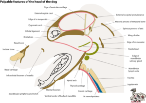
The head
Open the illustration in a new window or a new tab
Dorsal neck:
- Locate the spinous process of the axis, and cranially and laterally to this, the transverse processes of the atlas on each side. Determine the extent of rotation in the atlantoaxial joint with reference to these landmarks.
- Locate the external occipital protuberance. Determine the degree of flexion and extension possible in the occipitoatlantal joint with reference to this and the transverse processes of the atlas. These three landmarks are used to determine the placing of a needle for cerebrospinal; fluid collection from the cerebellomedullary cistern.
Dorsal head
- Palpate the external occipital protuberance and rostrally along the medial occipital crest medially between the muscle masses of m temporalis on each side.
- Locate the mastoid process of the petrous temporal bone near the external auditory meatus. At this level, the zygomatic arch leads rostrally to the orbit. The rim of the orbit is bony, except caudally where it is completed by the orbital ligament. This ligament, however feels hard and indistinguishable from bone.
- Over the bony surface of the maxilla, palpate the infraorbital vessels and nerve, and caudally to where they emerge from the infraorbital foramen.
- The rim of the osseous nasal opening is formed by the incisive bone laterally and the nasal bone dorsally. The nasal cartilages form the sides of the nasal vestibule.
Ventral neck
- Palpate the tracheal rings by pushing aside the covering ventrally of m sternohyoideus.
- Cranially palpate the arch of the cricoid cartilage, then to the ventrally protruding part of the thyroid cartilage.
- Rostral to this is the basihyoid bone. Pressing this should promote a swalowing movement during which the hyoid arch is protracted.
Ventral head
- Lateral to this are the large muscle masses of mm masseter and pterygoid fusing ventral to the body of the mandible. The parotid duct crosses the lateral surface of m masseter.
- Locate the mandibular salivary gland and mandibular lymph nodes medial to M pterygoideus.
- Rostral to the rostral edge of m masseter, the bony ventral border of the mandible is palpable. The mandibles meet medially at a mandibular notch ventrally. Caudal to this, locate the mental foramen.
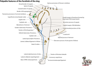
The forelimb
Open the illustration in a new window or a new tab
Scapula
- Dorsally, palpate the spinous processes of the thoracic vertebrae medially.
- Palpate the dorsal borders of right and left scapulae while moving these bones relative to the trunk.
- Palpate the spine of the scapula ventrally as far as the acromion.
- Cranial to the spine of the scapula, the muscle mass is formed by m supraspinatus.
- Move the scapula and locate the superficial cervical lymph deep to m omotransversarius.
Shoulder joint
- Locate the space between the acromion and the greater tuberosity of the humerus. Assess movements of the joint (flexion-extension, adduction-abduction, rotation, no gliding)
- Distally from the greater tuberosity, palpate the bony ridge of the deltoid crest.
- Locate the tendon of insertion of m intraspinatus as it passes over the lateral part of the greater tuberosity.
- Protract and retract the limb to assess the location of the pivot of the forelimb with the trunk, and to assess the contribution of the shoulder joint to this movement.
Thorax
- Locate the transverse processes of the cervical vertebrae and assess the location of the vertebral canal and spinal cord at the cranial end of the thorax.
- Locate the manubrium of the sternum and thereby the location and extent of the thoracic inlet.
- Locate the jugular groove. Compression of the veins caudally in the neck will occlude both the jugular vein and the cephalic vein that lies subcutaneously over the shoulder, laterally in the brachial region and cranially in the antebrachial region.
- Locate the costal arch and the xiphoid process of the sternum. Determine to location of the costochondral junctions relative to the costal arch.
Elbow joint
- The caudal border of the long head of m triceps leads to the olecranon process of the ulna. However, this landmark does not indicate the location of the elbow joint.
- Locate the firm fibrous band of the lateral collateral ligament of the elbow joint, and its attachment proximally to the lateral epicondyle of the humerus and distally to the head of the radius.
- Assess the normal movements of the elbow joint (flexion-extension, rotation, no adduction and abduction or gliding). Determine to what extent supination of the manus is possible when the elbow joint rotates. Compare observations on the dog with your own elbow.
Carpal joint
- Flex and extend the carpal joint to determine the location of the distal end of the ulna, the lateral styloid process, and the proximal end of the 5th metacarpal bone. Abduction of the joint is prevented by the lateral collateral ligement.
- Locate the accessory carpal bone and compare its mobility with the carpal joint flexed and extended. With the joint extended, feel the tension in the tendon attaching to it proximally, (m ulnaris lateralis) and the ligament attaching to it distally (accessoriometacarpal ligament). These are part of the mechanism preventing overextension (dorsal flexion) of the carpus and enabling the dog to be digitigrade.
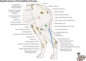
The hindlimb
Open the illustration in a new window or a new tab
Dorsal
- Palpate the spinous and transverse processes of the lumbar verebrae. Locate the crests of the ilia curving on each side. Further caudally, palpate the spinous processes of the sacrum as a fused median sacral crest.
- Dorsi- and ventriflex the tail to locate the most proximal moveable intervertebral joint, between the 1st and 2nd caudal vertbrae.
- Locate the ischiatic tuberosities and cranial to these, the sacrotuberous ligaments on each side of the pelvic cavity.
Hip joint
- Locate the greater trochanter of the femur. This and the ischiatic tuberosity are used to assess the movements of the hip joint. Push a finger between them and alternately flex and extend, abduct and adduct, and rotate the hip, while holding the dog in such a way as to ensure that only one hip joint is being moved. Only gliding is not possible.
Medial thigh
- Locate the prominent insertion of m pectineus on to the pubis. Press a finger into the depression cranial to this to feel the pulse of the femoral artery. This is the most convenient place to assess the pulse of the dog.
Stifle joint
- Compare the lateral and medial mobility of the patella with the stifle flexed and extended. With the stifle extended, it is easier to palate the medial and lateral ridges of the femoral trochlea. Also palpate the stifle joint capsule deep to the patellar ligament, and the insertion of the patellar ligament on to the tibial tuberosity at these two positions.
- Locate the medial and lateral collateral ligaments of the stifle, connecting the femoral epicondyles with the fibula laterally and the tibia medially.
- Also associated with the distal end of the femur, locate the gastrocnemius sesamoid bones on each side.
- Attempt adduction and adduction of the joint and assess what is preventing this.
- Ascertain that a small degree of rotation is possible. Ensure that you eliminate hip rotation from this assessment.
- Try to glide the tibia cranially and caudally with respect to the femur. This procedure assesses the integrity of the cranial and caudal cruciate ligaments respectively.
- Locate the popliteal fossa between mm biceps femoris and semitendinosus. The popliteal lymph node is palpable within this fossa if enlarged.
Hock joint
- Locate the insertion of the common calcanean tendon on to the calcanean tuberosity. Medial and cranial to the tendon, locate the tibial nerve.
- Locate the medial and lateral malleoli at the distal ends of the tibia and fibula respectively, and the associated medial and lateral collateral ligaments inserting on to the bases of the 2nd and 5th metacarpal bones.
- Flex the hock and palpate the medial tendon of insertion of m tibialis cranialis as it passes dorsally over the hock joint. This tendon is not palpable with the stifle extended. Deep to it, palpate the capsule of the talotibial joint. Differentiate this soft swelling with that of the lateral saphenous vein which is close by.
- Flex and extend the hock joint and note that the joint surfaces are angled so that the pes is abducted as the hock is extended.
- Attempt adduction and abduction, rotation and gliding, none of which is possible.
References
Ellenberger/Baum/Dittrich 1925 Labelled illustration reproduced in Nickel R, Schummer A, Seiferle E, Wilkens H, Wille K-H, Frewein J 1986 The locomotor system of the domestic mammals Paul Parey Berlin
Seiferle E 1952 Angewandte Anatomie am Lebenden Schweizer Archiv für Tierheilkunde 94 280-286
de Lahunta A Habel RE 1986 Applied veterinary anatomy WB Saunders Philadelphia
Goody PC 1997 Dog anatomy: a pictorial approach to canine structure JA Allen London
| This article has been peer reviewed but is awaiting expert review. If you would like to help with this, please see more information about expert reviewing. |
Error in widget FBRecommend: unable to write file /var/www/wikivet.net/extensions/Widgets/compiled_templates/wrt69a21290263d95_33374234 Error in widget google+: unable to write file /var/www/wikivet.net/extensions/Widgets/compiled_templates/wrt69a212902ad3b9_48998724 Error in widget TwitterTweet: unable to write file /var/www/wikivet.net/extensions/Widgets/compiled_templates/wrt69a21290307f62_57011889
|
| WikiVet® Introduction - Help WikiVet - Report a Problem |


