Exotic Urinary System - Anatomy & Physiology
Introduction
This section is devoted specifically to the renal anatomy and physiology of fish, aquatic and terrestrial amphibians, birds and reptiles. These animals excrete nitrogenous waste differently to domestic mammals, this combined with the very different habitats where these animals exist results in a variety of different renal mechanisms and appearances.
Nitrogenous Waste
Different organisms excrete nitrogen in different forms
Ammonia
- Some organisms excrete nitrogen directly as ammonia
- They are known as ammonotelic organisms
- This is how fish and aquatic amphibians excrete nitrogenous waste
- Relatively toxic but it is better tolerated by aquatic animals due to dilution
- 400ml of water is also excreted per gram of ammonia making this an unrealistic option for terrestrial organisms
Urea
- Some animals process ammonia into urea
- They are known as ureotelic organisms
- Mammals and terrestrial amphibians
- 40ml of water is excreted per gram of urea excreted
- Urea is very soluble and non-toxic
Uric Acid
- Referred to as Uricotelism
- Urea is further processed to uric acid
- Carried out by birds and reptiles
- Only 8ml of water is co-excreted per gram
- Highly insoluble and less toxic than ammonia, though it can precipitate into body cavities
Fish
Anatomy
Fish have a single kidney which is the same length as the coelom. It can be divided up into cranial and caudal parts; the cranial part has endocrine and haematopoietic functions and the caudal is where filtration occurs. It is not uncommon for some species to have no glomeruli however as a rule freshwater fish have larger glomeruli in greater numbers. Some species also have renal portal veins.
Osmoregulation
- Fish have no loop of henle and water movement is by osmosis
- Ammonia is removed via the urine and the gills
Freshwater
As the environment is hypotonic compared to the body of the fish ions are lost and water is gained across the gills therefore the kidney excretes water and has a very high glomerular filtration rate. The gills also undertake active uptake of NaCl and excrete ammonia and the diet is also very important for maintaining NaCl levels
Saltwater
The environment is hypotonic compared to the body of the fish therefore water is lost across their gills so they drink sea water to replace this which results in a large intake of salt (activates Angiotensin 2). They excrete both ammonia and NaCl across their gills and further NaCl across their skin. Their kidneys have small or absent glomeruli and their main function is the elimination of excess divalent ions e.g. Mg2+
Amphibian
Anatomy
In amphibians urine moves from the kidney down the ducts into the cloaca and then onto a urinary bladder. Caudates and Anurans possess renal portal veins which carry blood from the hind limbs to the kidney before it goes back to the heart. They have paired posterior kidneys which lie retroperitoneally. Caecilians have no renal portal veins and have one kidney the full length of the coelom
Physiology
- Aquatic amphibians excrete ammonia whereas terrestrial species excrete uric acid
- Their kidneys filter coelomic and or vascular fluid
- They have a high GFR
- Their urine is hypo-osmotic
- They have a urinary bladder
Aquatic Amphibians
Aquatic amphibians have extremely water permeable skin and therefore lots of osmosis occurs across the skin. It falls to the kidney to excrete excess water and ammonia
Terrestrial Amphibians
Terrestrial amphibians have the totally opposite problem to their aquatic counterparts. To them water conservation is very important as water is lost via various routes including evaporation. They therefore have a urinary bladder which is permeable and water is reabsorbed across it this is controlled by arginine vasotocin (AVT). This chemical increases the number of aquaporins in the membrane. They excrete urea and are able to decrease their GFR if water is reduced.
Avian
Anatomy
Birds have paired kidneys which account for 1 – 2.5% of their body weight which is significant compared to 0.5% in mammals. They are located caudal to the caudal edge of the lungs near the abdominal air sac diverticulum. They are subdivided into cranial, middle and caudal parts and have lobules comprising a cortex and a medullar cone. Birds have a renal portal system similar to that of reptiles.
Types of Nephron
There are two possible types of nephrons found in birds
- The first type is similar to that of reptiles
- No loop of henle
- Cortex only
- The second type is more similar to that of mammals and is found in 10 – 30% of species
- Loop of henle’s are present
- Cortex and medulla
- However both systems only allow for limited urine concentration
Physiology of the Elimination of Uric Acid
- Birds excrete uric acid as a white / light yellow colloidal suspension
- Uric acid crystals precipitate (no osmotic pressure)
- Small volume
- Precipitate contains uric acid, sodium/potassium and protein
- Urine enter cloaca and mixes with the faecal material
Salt glands
These glands are found in desert and aquatic birds as salt consumption exceeds renal clearance. These supraorbital glands drain into the nostrils and account for 20% of total NaCl excretion. They are not under the control of kidneys and they hypertrophy if the birds salt intake increases.
Avian Renal Portal System
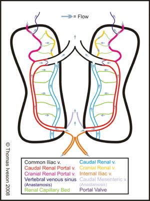
In birds the blood from the hindlimbs is carried directly to the kidneys. The cranial and caudal renal portal veins deliver blood from the hindlimbs into the capillary beds of the interlobar arteries. Therefore blood which has already been through capillary beds in the hind limbs mixes with blood directly from the heart. It bypasses the glomeruli and waste products in the blood are secreted directly into the tubules. This is thought to be a more efficent way to excrete uric acid and urate. A portal valve regulates how much blood from the hindlimbs passes through the kidneys. When open very little blood enters the capillary beds as it follows the path of least resistance. If the valve narrows then blood is forced into the capillary beds. However some blood always escapes thanks to connections with both the caudal mesenteric veins and vertebral sinuses. It is of significance when injecting these animals if the injection is given in the caudal half of the body most of the drug will be potentially lost in the urine before it has time to act it may also be toxic to the kidney as it has not been metabolised by the liver
Other Roles of the Avian Kidney
- Activates vitamin D
Reptile
Gross Renal Anatomy of Lizards
Two kidneys are present in lizards but the caudal aspect of them is fused in many species. Also the presence of a urinary bladder is species specific.
Gross Renal Anatomy of Snakes
- Snakes have paired kidneys with the right being most cranial.
- The kidneys are comprised of 25-30 lobules
- No bladder
- Urine is stored in either the distal colon or flared ends on each urethra
Gross Renal Anatomy of Chelonians
Chelonians have an osmotically permeable bladder which can reabsorb water. This structure acts as a buoyancy aid in aquatic turtles and helps reabsorb sodium. Some species have paired accessory bladders off the main structure
Microscopic Renal Anatomy of Reptiles
Reptiles have no pelvis, pyramids, cortex or medulla and kidney is only made up of a few thousand nephrons with poorly developed glomeruli. Few capillaries supply the kidneys and the nephrons have no loop of henle. In squamate males a sexual segment between the distal tubule and collecting ducts is present.
Nitrogenous Waste
Reptiles excrete nitrogenous waste mainly in the form of uric acid. It is suspended in spheres complexed with protein and sodium (carnivorous diet) or potassium (herbivorous diet) along with mucoid substances (glycoprotein and mucopolysaccharides). As a result their urine contains large quantities of protein.
Uric Acid Secretion in Reptiles
Uric acid is secreted into the proximal tubules actively using potassium and into the bladder (where present) is response to H+ secretion. The secretion of urate increases in response to a decrease in blood pH.
In the urodeum urine moves via reverse peristalsis to the rectum where some protein is reabsorbed to be recycled
Post Renal Urine Modification in Reptiles
Voided urine is not reflective of kidney function due to the transport of ions and water across the colon wall and the reabsorption of sodium / excretion of potassium and urates in the bladder.
Reptilian Renal Adaptations for Water Conservation
- Very few nephrons therefore low GFR
- They secrete uric acid
- Able to decrease GFR in times of stress
- Salt glands allow excretion of sodium and potassium without concurrent water loss
- Water is reabsorbed from urine in the colon
Reptilian Response to Dehydration
- The afferent arteriole collapses in response to increased levels of arginine vasotocin
- The glomeruli close and the tubules collapse
- This results in a significantly decreased GFR and therefore decreased excretion
- Renal portal blood perfuses the tubules
Reptilian Renal Portal System
- Similar to that of birds
- The renal portal vein bypasses the glomeruli of the kidneys
- In some species it has a valve
- If closed
- Blood goes from the hindlimbs to the kidneys to the heart
- Valves open
- Response to stress
- Blood bypasses kidney
- If closed
Significance
- This means if the animal is stressed and is injected in the caudal half of the body the drug will have greater effects as it will not be filtered
- If the animal is not stressed most of the drug will be potentially lost in the urine before it has time to act it may also be toxic to the kidney as it has not been metabolised by the liver
- This is why it is best to inject them in the cranial half of the body
Reptilian Salt Glands
- Similar to birds
- Actively secrete sodium and potassium
- They are located near the eye or nasal passages
- They are stimulated by a high plasma osmotic concentration
- Allow reptiles to “sneeze” excess salt
- Sea turtles have modified tear glands which allow them to secrete salt from their eyes
Other Roles of the Reptilian Kidney
- Activates vitamin D
- Synthesises Vitamin C
