Spinal Cord - Anatomy & Physiology
| This article is still under construction. |
Introduction
The spinal cord can be divided to several regions; cervical (C1-C6), cervicothoracic (C7-T2), thoracolumbar (T3-L3), lumbosacral (L3-S2) and sacral (S3 onwards). Nerves originating from the spinal cord and the segmental spinal nerves innervate the limbs.
The forelimb nerves include the suprascapular (C5-6), the musculocutaneous (C5-7), the ulna/median (Originates from the brachial plexus, which is formed from C5-T1) and the radial (C5-T1). The hindlimb nerves include the obturator (L2-4), the femoral (L2-4) and the sciatic (L4-S3). The sciatic nerve branches to the tibial nerve and the peroneal nerve.
Structure and Function
The spinal cord' is constructed of the marginal layerwhich has axons and white matter, the mantle which contains cell bodies and grey matter and the spinal canal. This canal conducts sensory information from the peripheral nervous system (both somatic and autonomic) to the brain, conducts motor information from the brain to various effectors and acts as a minor reflex center.
Function
Components
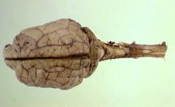
The central nervous system consists of; the brain (Prefix = "encephalo") and the spinal cord (Prefix = "myelo").
Function
Sensory neurons from both the internal and external environment relay information to the CNS. The CNS processes sensory information and intitiates motor outputs. Effector and motor neurons from the CNS relay the appropriate outputs to effector organs.
The Autonomic Nervous System
The autonomic nervous system relays sensory information from, and motor information to, the internal environment. It therefore plays an important role in the maintenance of homeostasis.
The Somatosensory Nervous System
The somatosensory nervous system relays sensory information from, and motor information to, the external environment.
White and Grey Matter
White Matter
White matter consists of accumulations of myelinated axons. Myelinated axons are wrapped in myelin. Myelin is compsed of lipid and protein in an 80:20 ratio. It insulates axons to give efficient action potential conduction. Myelin is provided by oligodendrocytes in the CNS. They respond poorly in injury. Schwann cells in the PNS myelinate one axon only. A "funiculus" is a large region of white matter in the spinal cord.
Grey Matter
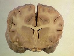
The outer portions of the cerebral cortex and the inner portions of the spinal cord are composed of grey matter. Grey matter is also found in coloumns and scattered in brainstem nuclei. It is composed of neuronal cell bodies, plus glial cells.
Upper and Lower Motor Neurons
Lower Motor Neuron (LMN)
LMNs are efferent neurons which connect the CNS to smooth or skeletal muscle. Autonomic LMNs connect to smooth muscle. Somatic LMNs connect to skeletal muscle. Those innervating the muscles of the axial and peripheral skeleton have their cells bodies in the ventral horn of the spinal cord. Injury causes LMN weakness. This is characterised by; Depressed reflexes, decreased tone and neurogenic muscle atrophy.
Upper Motor Neuron (UMN)
The upper motor neuron comprises the motor system of the CNS. This is responsible for; Initiating voluntary movement and maintenance of tone and posture. In man, direct connections exist between neurons in the motor cortex and LMNs in the spinal cord. This is known as the "pyramidal system". In animals, there are scattered groups of interconnected neurons in the cortex and brainstem, which ultimately synapse with LMNs in the brainstem and spinal cord. This is called the "extrapyramidal system". UMN injury results in increased extensor tone, giving; Stiffness, spasticity, delay in the onset of protraction, and a longer stride, disinhibition of the LMN relfex ability, causing increased reflexes, the inability to stimulate the LMN and UMN weakness results.

