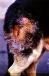
Bacterial skin infections - Pathology
Cutaneous bacterial infections tend to be called pyodermas. They are superficial, deep and are common in dogs, but less common in other species.
Superficial Pyoderma
- Affects epidermis and upper infundibulum of hair follicles
- No scarring when healed
- Grossly:
- Erythema
- Alopecia
- Papules and pustules
- Crusts
- Epidermal collarettes
- Microscopically:
- Intraepidermal pustular dermatitis
- Superficial suppurative folliculitis
- Bacteria commonly not seen
Impetigo
Dermatophilosis
Greasy pig disease
Ovine Fleece Rot
Equine Pastern Folliculitis
Deep pyoderma
- Less common than superficial pyoderma
- Occurs mainly in dogs
- Affects infundibulum, isthmic portion of hair follicles and surrounding dermis and subcutis
- Heals with scarring
- Local lymph nodes are often affected
- Often secondary to immunosuppression, follicular hyperkeratosis or demodicosis
- May also be a sequele to superficial pyoderma
- Grossly:
- Microscopically:
- Pyogranulomatous folliculitis and furunculosis
- Nodular or diffuse dermatitis
- Panniculitis
- May involve a foreign bodey reaction to follicular contents and draining sinuses develop
- If chronic, scarring and loss of adnexa
- Bacteria often isolated include Staphylococcus spp., especially S. intermedius in dogs, Streptococcus spp., Corynebacterium pseudotuberculosis, Pseudomonas, Pasteurella, Proteus, E.coli
Staphylococcal Folliculitis and Furunculosis
Subcutaneous Abscesses
- Purulent exudate within dermis and subcutis
- Commonly occurs in cats due to contamination of penetrating wounds
- Surrounding wall of collagen and fibroblasts may develop
- Common bacteria (often normal mouth flora)
Bacterial Granulomatous Dermatitis
- Usually due to saprophytes
- Grossly:
- Diffuse or nodular lesions
- May ulcerate and form drainage fistulas
- Microscopically:
- Macrophages +/- multinucleated giant cells
- Caseous necrosis and neutrophils
- Mycobacterial granulomatous or pyogranulomatous lesions
- Usually caused by Mycobacterium lepraemurium (feline leprosy) or other Mycobacteria
- Most commonly lesions appear on head, neck and legs
- Botryomycosis
Bacterial Pododermatitis
- Digital infections in ruminants
- Contagious Footrot
- Necrobacillosis of the foot
Systemic Bacterial Infections
- Salmonellosis
- Capillary dilatation and congestion -> cyanosis of external ears and abdoman
- Thrombosis -> necrosis of extremities
- Erysipelas in pigs
- Caused by Erysipelothrix rhusiopathiae
- Vasculitis, thrombosis, ischaemia -> cutaneous lesions - firm, raises, rhomboidal pink to dark purple areas
- Clostridium novyi
- Severe cellulitis, toxaemia and death of young rams during breeding season (due to traumatised heads) - 'big head'
- Streptococcus equi
- In horses
- Immune complex vasculitis -> purpura
